Home > Science > SEM
Tapeworm, SEM C016 / 9076

![]()
Wall Art and Photo Gifts from Science Photo Library
Tapeworm, SEM C016 / 9076
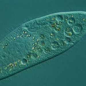
Tapeworm. Coloured scanning electron micrograph (SEM) of a tapeworm (Taenia pisiformis), showing the hooks (upper right) it uses to cling to its host. Tapeworms are parasitic flatworms that live in the digestive tract of their vertebrate host. T. pisiformis is one of the most common parasites of the dog (the other being Dipylidium caninum). It is found worldwide. Magnification: x100, when printed 10 centimetres wide
Science Photo Library features Science and Medical images including photos and illustrations
Media ID 9244625
© STEVE GSCHMEISSNER/SCIENCE PHOTO LIBRARY
Canine Cestode Colored Cyclophyllid Electron Microscope Flatworm Hook Hooks Parasite Parasitic Parasitism Parasitological Parasitology Scolex Taenia Pisiformis Tapeworm
EDITORS COMMENTS
This print showcases the intricate world of a tapeworm, providing a close-up view of its fascinating anatomy. The coloured scanning electron micrograph (SEM) reveals the hooks that this parasitic flatworm employs to firmly attach itself to its host. With an impressive magnification of x100, every detail is brought to life on the white background. Tapeworms like Taenia pisiformis are notorious parasites that inhabit the digestive tract of vertebrate hosts. In particular, T. pisiformis is one of the most prevalent parasites found in dogs worldwide, alongside Dipylidium caninum. This image sheds light on their presence and prevalence in these animals. The SEM technique used here allows us to appreciate the complexity and beauty within nature's smallest creatures. By capturing this microscopic snapshot through an electron microscope, we gain insight into how these hooks aid tapeworms in their survival and reproduction. Beyond its scientific significance, this photograph serves as a reminder of our interconnectedness with wildlife and highlights the diverse range of organisms coexisting with us on Earth. It invites contemplation about our role as stewards of nature and prompts curiosity about other unseen wonders lurking beneath our awareness. Photographer Steve Gschmeissner skillfully captures both artistry and science in this print from Science Photo Library, offering viewers a glimpse into a hidden realm where biology meets aesthetics seamlessly.
MADE IN AUSTRALIA
Safe Shipping with 30 Day Money Back Guarantee
FREE PERSONALISATION*
We are proud to offer a range of customisation features including Personalised Captions, Color Filters and Picture Zoom Tools
SECURE PAYMENTS
We happily accept a wide range of payment options so you can pay for the things you need in the way that is most convenient for you
* Options may vary by product and licensing agreement. Zoomed Pictures can be adjusted in the Cart.