
Protein Collection
Protein: The Building Blocks of Life Proteins, the fundamental components of cells, play a crucial role in maintaining our body's functions


All Professionally Made to Order for Quick Shipping
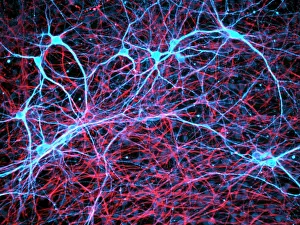
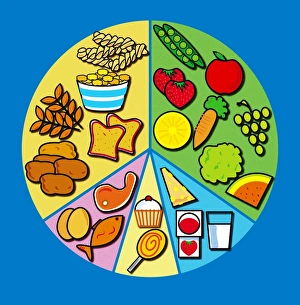
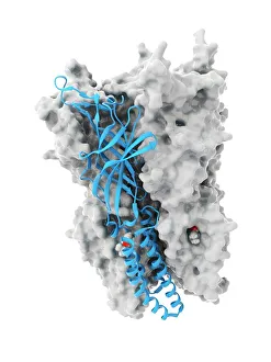
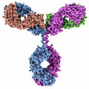
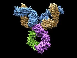
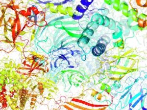
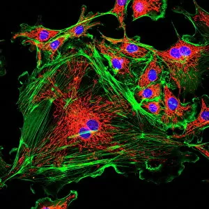
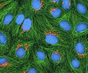
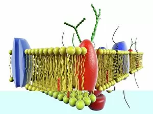
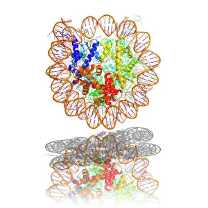
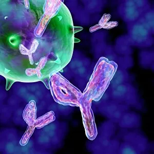
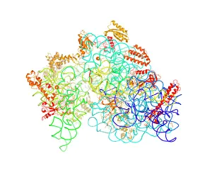
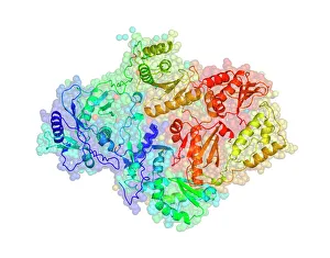
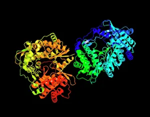
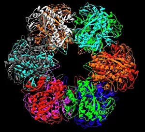
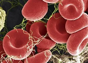
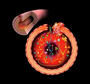
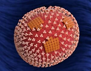
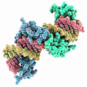
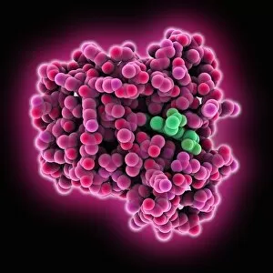
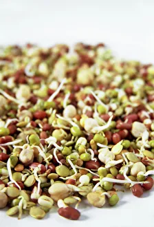
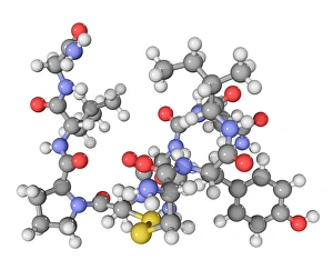
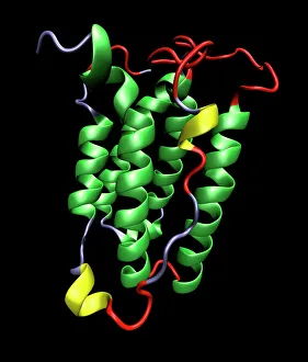
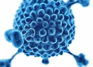
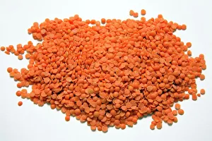
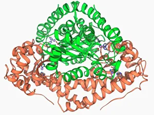
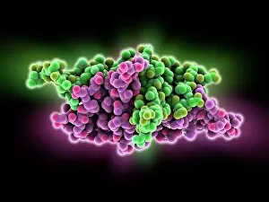
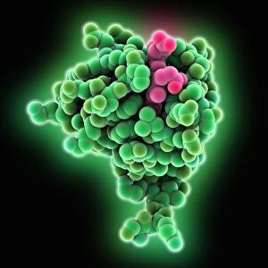
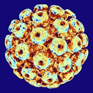
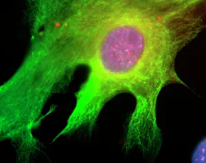
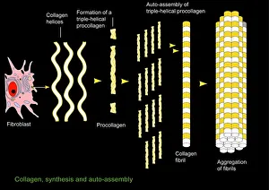
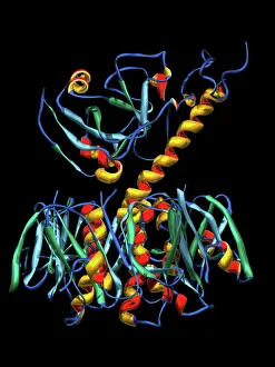
Protein: The Building Blocks of Life Proteins, the fundamental components of cells, play a crucial role in maintaining our body's functions. From nerve and glial cells to DNA transcription they can involved in every aspect of life. In a mesmerizing light micrograph, we witness the intricate network formed by nerve and glial cells. These proteins ensure proper communication between neurons and support their growth and survival. An anaesthetic inhibiting an ion channel is captured in another captivating image (C015 / 6718). This protein acts as a gatekeeper for ions entering or leaving the cell, controlling its electrical activity. DNA transcription is depicted through a molecular model. Proteins called RNA polymerases read our genetic information and synthesize messenger RNA molecules that carry instructions for building other proteins. Glial cells take center stage once again in a confocal light micrograph. These supportive proteins protect neurons from damage while also regulating their environment to maintain optimal functioning. HeLa cells shine brightly under the microscope (C017 / 8299). Derived from cervical cancer tissue, these immortalized human cells have contributed immensely to scientific discoveries involving protein research. The immunoglobulin G antibody molecule stands tall as it fights against pathogens invading our bodies. This remarkable protein recognizes foreign substances and aids in neutralizing them (F007 / 9894). Away from cellular wonders, doner kebab cooking reminds us that even delicious food contains ample amounts of protein. Istanbul showcases this culinary delight which has become popular across Europe. Maintaining a balanced diet ensures sufficient intake of essential amino acids found abundantly in various protein sources like meat, fish, legumes, dairy products, nuts, and seeds. Artwork depicting metabolic enzymes highlights how these specialized proteins drive chemical reactions within our bodies' metabolism – converting nutrients into energy or building blocks for growth and repair. The secondary structure of proteins comes alive through artistic representation.