
Receptors Collection
Receptors: Unlocking the Secrets of Sensation From anaesthetic inhibiting ion channels to brain protein research


All Professionally Made to Order for Quick Shipping
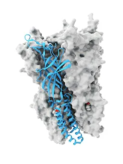
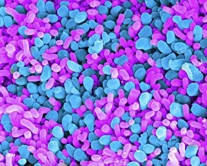
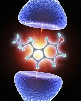
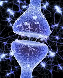
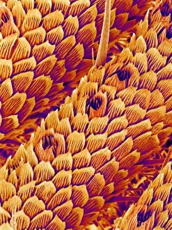
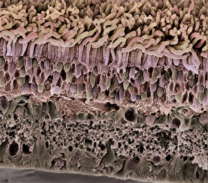
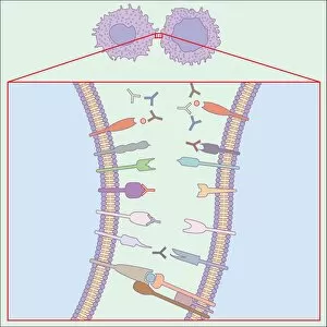
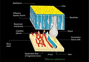
Receptors: Unlocking the Secrets of Sensation From anaesthetic inhibiting ion channels to brain protein research, receptors play a crucial role in our understanding of sensory perception. These intricate structures are like gatekeepers, allowing signals to pass through and enabling us to experience the world around us. In the realm of touch, touch receptors act as messengers between our skin and brain, relaying information about texture, pressure, and temperature. They allow us to feel the gentle caress of a loved one or warn us when something is too hot to handle. Our eyesight relies on rod and cone cells found in the retina. Through scanning electron microscopy (SEM), we can marvel at their intricate structure that enables us to see colors and shapes with astonishing clarity. The rods work tirelessly in low light conditions while cones bring vibrant hues into focus during daylight. The human olfactory system is another fascinating area where receptors come into play. With artwork depicting this complex network, we gain insight into how odor molecules interact with receptor proteins in our noses, triggering memories or alerting us to danger. Nerve synapses serve as vital connections within our nervous system. Artwork showcasing these junctions reveals their complexity - transmitting electrical impulses from one neuron to another with lightning speed. Understanding these synapses helps unravel mysteries surrounding learning, memory formation, and even mental health disorders. As researchers delve deeper into receptor function and regulation mechanisms such as anaesthetics inhibiting ion channels or investigating brain proteins' roles, new breakthroughs emerge daily. Picture No. 10877011 captures a moment frozen in time - an image that represents countless hours spent uncovering nature's secrets. Receptors hold immense potential for medical advancements; they offer hope for developing targeted therapies for various diseases affecting sensation pathways or improving anesthesia techniques by precisely targeting specific ion channels. Intricate yet awe-inspiring, receptors continue captivating scientists worldwide as they strive towards unraveling the complexities of human perception.