Fraud Blocker Skip to main content

Reproductive Part Collection

"Exploring the Intricate World of Reproductive Parts

Background imageReproductive Part Collection: English oak (Quercus robur) acorns

English oak (Quercus robur) acorns. Acorns forming on an English oak tree. This tree is also known as the pedunculate oak. Acorns are the fruit of the oak tree

Background imageReproductive Part Collection: Passion flower pollen, SEM

Passion flower pollen, SEM
Passion flower pollen. Coloured scanning electron micrograph (SEM) of a passion flower (Passiflora caerulea) pollen grain. Pollen grains are the male gametes (sex cells) of a plant

Background imageReproductive Part Collection: Dandelion pollen grain, SEM

Dandelion pollen grain, SEM
Dandelion (Taraxacum officinale) pollen grain, coloured scanning electron micrograph (SEM). Pollen grains are reproductive structures produced by the male parts of flowering plants

Background imageReproductive Part Collection: Philadelphia fleabane pollen grains, SEM

Philadelphia fleabane pollen grains, SEM
Philadelphia fleabane (Erigeron philadelphicus) pollen grains, coloured scanning electron micrograph (SEM). Pollen grains are reproductive structures produced by the male parts of flowering plants

Background imageReproductive Part Collection: Gorse stigma with pollen grains, SEM

Gorse stigma with pollen grains, SEM
Gorse (Ulex europaeus) stigma (green) with pollen grains (yellow), coloured scanning electron micrograph (SEM). Pollen grains are reproductive structures produced by the male parts of flowering

Background imageReproductive Part Collection: Lily pollen grain on rosemary leaf, SEM

Lily pollen grain on rosemary leaf, SEM
Lily pollen grain on rosemary leaf. Coloured scanning electron micrograph (SEM) of a pollen grain (yellow) from a lily flower (Lilium sp.)

Background imageReproductive Part Collection: Forsythia pollen grains, SEM

Forsythia pollen grains, SEM
Forsythia pollen grains arranged in a spiral, coloured scanning electron micrograph (SEM). Pollen grains are reproductive structures produced by the male parts of flowering plants

Background imageReproductive Part Collection: Chickweed pollen grains, SEM

Chickweed pollen grains, SEM
Chickweed pollen grains. Coloured scanning electron micrograph (SEM) of pollen grains (green) from a chickweed flower (Stellaria media)

Background imageReproductive Part Collection: Chickweed pollen grain, SEM

Chickweed pollen grain, SEM
Chickweed pollen grain. Coloured scanning electron micrograph (SEM) of a pollen grain (green) from a chickweed flower (Stellaria media)

Background imageReproductive Part Collection: Red Campion Seed (Silene dioica) SEM

Red Campion Seed (Silene dioica) SEM
Red campion seed. Coloured scanning electron micrograph (SEM) of a seed from a red campion plant (Silene dioica). Seed shapes

Background imageReproductive Part Collection: Basil pollen grain, SEM

Basil pollen grain, SEM
Basil pollen grain. Coloured scanning electron micrograph (SEM) of a pollen grain from the flower of a basil (Ocimum basilicum) plant. Pollen grains are the male gametes (sex cells) of a plant

Background imageReproductive Part Collection: Fungal spores on pollen grain, SEM

Fungal spores on pollen grain, SEM
Fungal spores on pollen grain. Coloured scanning electron micrograph (SEM) showing fungal spores (round) on a pollen grain from a goji berry (wolfberry, Lycium sp.) plant

Background imageReproductive Part Collection: Dodder seed, SEM C015 / 5077

Dodder seed, SEM C015 / 5077
Dodder (Cuscuta sp.) sede, coloured environmental scanning electron micrograph (ESEM)

Background imageReproductive Part Collection: Grey Willow (Salix cinerea) C014 / 0492

Grey Willow (Salix cinerea) C014 / 0492
Grey Willow (Salix cinerea) flowering in Spring, showing male catkins with pollen

Background imageReproductive Part Collection: Grey Willow (Salix cinerea) C014 / 0491

Grey Willow (Salix cinerea) C014 / 0491
Grey Willow (Salix cinerea) flowering in Spring, showing a male catkin with pollen

Background imageReproductive Part Collection: Campion Seed Detail (Silene dioica) SEM

Campion Seed Detail (Silene dioica) SEM
Red campion seed detail. Coloured scanning electron micrograph (SEM) of part of a seed from a red campion plant (Silene dioica)

Background imageReproductive Part Collection: Aubergine pollen grain, SEM

Aubergine pollen grain, SEM
Aubergine pollen grain. Coloured scanning electron micrograph (SEM) of a pollen grain from the flower of an aubergine (Solanum melongena, eggplant) plant

Background imageReproductive Part Collection: Hyssop pollen grains, SEM

Hyssop pollen grains, SEM
Hyssop pollen grains. Coloured scanning electron micrograph (SEM) of pollen grains (blue-green) from the herb hyssop (Hyssopus officinalis) plant on a petal (red)

Background imageReproductive Part Collection: Scots pine pollen, SEM

Scots pine pollen, SEM
Scots pine pollen. Coloured scanning electron micrograph (SEM) of Scots pine (Pinus sylvestris) pollen grains. The pollen grains consist of a base (orange) with two air-filled

Background imageReproductive Part Collection: Scots pine pollen grain, SEM

Scots pine pollen grain, SEM
Scots pine (Pinus sylvestris) pollen grain, coloured scanning electron micrograph (SEM). The pollen grain has two air-filled, wing-like bladders (pink)

Background imageReproductive Part Collection: Chinese hibiscus pollen, SEM

Chinese hibiscus pollen, SEM
Chinese hibiscus pollen. Coloured scanning electron micrograph (SEM) of a pollen grain from a Chinese hibiscus (Hibiscus rosa-sinensis) flower

Background imageReproductive Part Collection: Bellflower pollen, SEM

Bellflower pollen, SEM
Bellflower pollen. Coloured scanning electron micrograph (SEM) of pollen grains from a bellflower (Campanula sp.). Pollen grains are the male gametes (sex cells) of a plant

Background imageReproductive Part Collection: Tulip (Tulipa gesneriana), hybrid form C010 / 7011

Tulip (Tulipa gesneriana), hybrid form C010 / 7011
Tulip (Tulipa gesneriana) flowering in Spring, showing the stigma and stamens

Background imageReproductive Part Collection: Mirabilis anthers, light micrograph

Mirabilis anthers, light micrograph. These anthers, seen from the side, are from Mirabilis jalapa, the four o clock flower. The anthers and the filament (lower right) together form a stamen

Background imageReproductive Part Collection: Mirabilis anther, light micrograph

Mirabilis anther, light micrograph
Mirabilis anther and pollen grains, confocal fluorescence light micrograph. This anther is from Mirabilis jalapa, the four o clock flower

Background imageReproductive Part Collection: Orbea ciliata flower

Orbea ciliata flower. Close-up of the reproductive organs of the succulent Orbea ciliata. Photographed in Cape Province, South Africa, in July

Background imageReproductive Part Collection: Celandine (Chelidonium majus) seeds

Celandine (Chelidonium majus) seeds
Greater celandine (Chelidonium majus) seeds. Close-up of seeds from the Greater celandine (Chelidonium majus) plant, showing elaiosomes (white)

Background imageReproductive Part Collection: Passion flower pollen, SEM

Passion flower pollen, SEM
Passion flower pollen. Coloured scanning electron micrograph (SEM) of pollen grains from a passion flower (Passiflora caerulea). Pollen grains are the male gametes (sex cells) of a plant

Background imageReproductive Part Collection: Lily reproductive parts

Lily reproductive parts. Close-up of the stamens and carpel of a hybrid garden lily. Lilies display three-fold symmetry in their floral parts

Background imageReproductive Part Collection: Dandelion pollen, SEM

Dandelion pollen, SEM
Dandelion (Taraxacum officinale) pollen. Coloured scanning electron micrograph (SEM) showing pollen grains (round) on the anther of a dandelion flower

Background imageReproductive Part Collection: Geranium magnificum in flower

Geranium magnificum in flower. Photographed in Dorset, UK, in July

Background imageReproductive Part Collection: European larch cones

European larch cones
European larch (Larix decidua) cones forming in spring. Photographed in April, in Dorset, UK

Background imageReproductive Part Collection: Dandelion pollen grains, SEM

Dandelion pollen grains, SEM
Dandelion (Taraxacum officinale) pollen grains, coloured scanning electron micrograph (SEM). Pollen grains are reproductive structures produced by the male parts of flowering plants

Background imageReproductive Part Collection: Geranium Blue cloud in flower

Geranium Blue cloud in flower. Photographed in Dorset, UK, in July

Background imageReproductive Part Collection: Thale cress anther and pollen, micrograph

Thale cress anther and pollen, micrograph
Thale cress anther and pollenr. Fluorescence micrograph of an anther (male reproductive structure) from a thale cress (Arabidopsis thaliana) flower, showing the pollen grains (round) inside

Background imageReproductive Part Collection: Thale cress stamens, micrograph

Thale cress stamens, micrograph
Mitosis. Confocal light micrograph of the stages mitosis (nuclear division) and cytokinesis (cell division). During mitosis the nuclear envelope disintegrates (3rd image) and the chromosomes (purple)

Background imageReproductive Part Collection: Thale cress flower, micrograph

Thale cress flower, micrograph
Thale cress flower. Fluorescence micrograph of a flower from a thale cress (Arabidopsis thaliana) plant. The female reproductive structures - carpel (red) and stigma (yellow) - can be seen at centre

Background imageReproductive Part Collection: Thale cress anther, micrograph

Thale cress anther, micrograph
Thale cress anther. Fluorescence micrograph of an anther (male reproductive structure) from a thale cress (Arabidopsis thaliana) flower, showing the pollen grains (red, round) inside

Background imageReproductive Part Collection: Thale cress stigma, micrograph

Thale cress stigma, micrograph
Thale cress stigma. Fluorescence micrograph of a stigma (female reproductive structure) from a thale cress (Arabidopsis thaliana) flower

Background imageReproductive Part Collection: Stamens and carpels, micrograph

Stamens and carpels, micrograph
Stamens and carpels. Fluorescence micrograph of stamens (male reproductive structures, far left) and carpels (female reproductive structures)

Background imageReproductive Part Collection: Thale cress stigma, micrograph

Thale cress stigma, micrograph
Thale cress stigma. Fluorescence micrograph of a stigma (female reproductive structure) from a thale cress (Arabidopsis thaliana) flower

Background imageReproductive Part Collection: Meadowsweet, artwork

Meadowsweet, artwork
Meadowsweet (Filipendula ulmaria), artwork. This plant is a herb that grows in damp meadows in most of Europe and in western Asia

Background imageReproductive Part Collection: Thorn apple (Datura stramonium) seed pod

Thorn apple (Datura stramonium) seed pod. This plant is a member of the nightshade family. It contains poisonous alkaloids. Photographed in disturbed ground in the UK

Background imageReproductive Part Collection: Common snowdrop, artwork

Common snowdrop, artwork
Common snowdrop (Galanthus nivalis), artwork. This flowering plant is found throughout Europe. Its white flower is pendulous (hangs downwards), and it is the among the first flowers to bloom in spring

Background imageReproductive Part Collection: Common primrose, artwork

Common primrose, artwork
Common primrose (Primula vulgaris), artwork. This flowering plant is found in temperate Europe and further south and west into Turkey and Iran

Background imageReproductive Part Collection: Crimson waxcap mushrooms, artwork

Crimson waxcap mushrooms, artwork
Crimson waxcap (Hygrocybe punicea) mushrooms, artwork. This fungus is found in northern Europe. The mushrooms (fruiting bodies) have a bell-shaped cap (lower right)

Background imageReproductive Part Collection: Lesser celandine, artwork

Lesser celandine, artwork
Lesser celandine (Ranunculus ficaria), artwork. This flowering plant is found in Europe and western Asia, mostly on damp ground

Background imageReproductive Part Collection: Aubergine (Solanum melongena) flower

Aubergine (Solanum melongena) flower. Close-up of a flower on an aubergine plant. Photographed in Orvieto, Umbria, Italy


All Professionally Made to Order for Quick Shipping

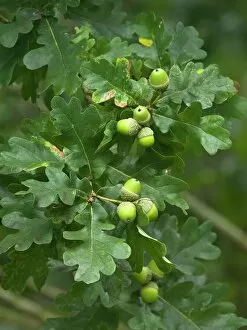
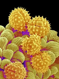
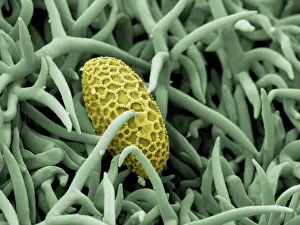
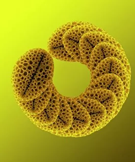
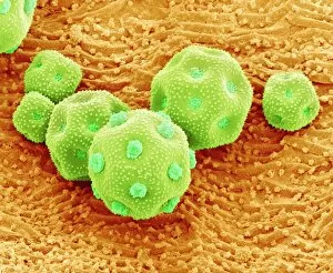
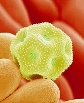
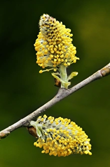
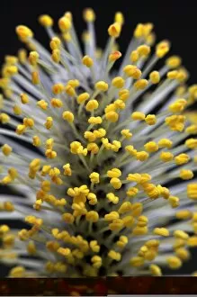
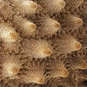
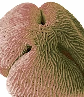
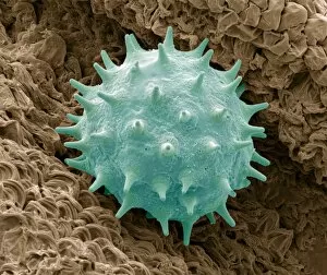
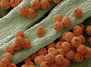
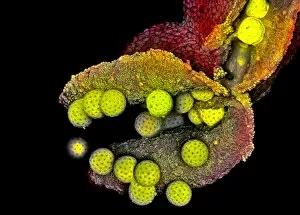
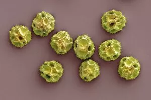
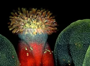
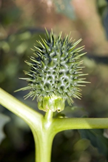
"Exploring the Intricate World of Reproductive Parts: A Microscopic Journey" Embark on a fascinating journey into the hidden realm of reproductive parts as we delve into the captivating beauty and intricate structures of various plant species. From English oak acorns to delicate passion flower pollen, each image captured under a scanning electron microscope (SEM) unveils nature's remarkable mechanisms for reproduction. Witness the mesmerizing patterns and textures present in dandelion pollen grains, resembling tiny spheres adorned with delicate spikes. Philadelphia fleabane pollen grains showcase their unique shape and surface features, while gorse stigma reveals its vibrant yellow hue along with an abundance of attached pollen grains. A striking contrast emerges when lily pollen grain finds its temporary resting place on a rosemary leaf, creating an unexpected juxtaposition between two distinct botanical elements. Forsythia's bright yellow petals give way to its SEM-captured pollen grains that appear like miniature golden orbs suspended in mid-air. Marvel at chickweed's minuscule yet intricately designed pollen grains, showcasing exquisite details that are often overlooked by the naked eye. Passion flower takes center stage once again as its SEM image highlights the complex structure and interwoven threads within each individual grain. Chinese hibiscus surprises us with its vibrant red coloration and distinctive spherical-shaped pollens that seem almost otherworldly under high magnification. Bellflower adds another layer of diversity to our exploration with its own unique set of SEM-captured pollens boasting intriguing shapes and textures. Finally, we encounter thale cress flower micrograph – a stunning portrayal capturing this small flowering plant's reproductive part in all its glory. Delve deeper into this microscopic world where every detail holds significance in ensuring successful reproduction for these remarkable plants. Through these captivating images taken through powerful microscopy techniques, we gain a newfound appreciation for nature's ingenuity in propagating life itself – reminding us how even the tiniest components play a crucial role in the grand tapestry of existence.