
Retrovirus Collection
"Unveiling the Intricacies of Retroviruses: A Microscopic Journey into HIV Infection" In this captivating collection of images


All Professionally Made to Order for Quick Shipping
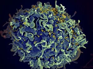
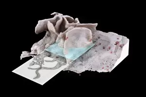
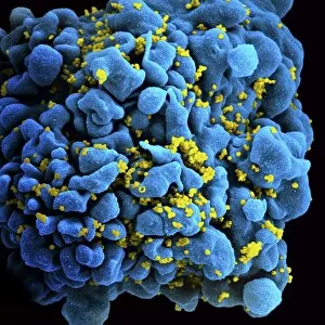
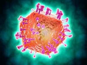
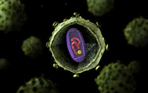
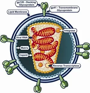
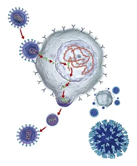
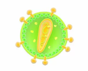
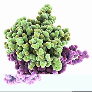
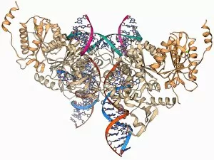
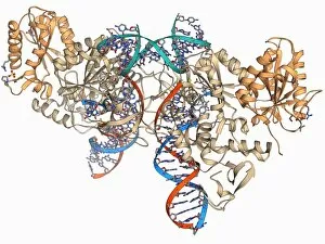
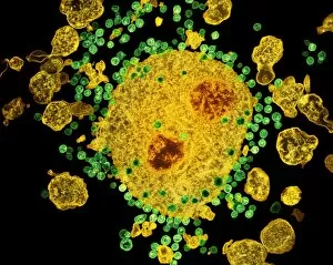
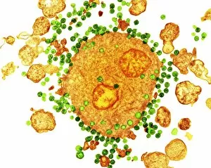
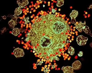
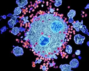
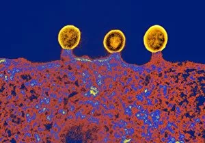
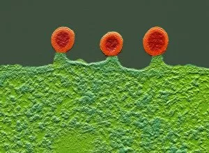
"Unveiling the Intricacies of Retroviruses: A Microscopic Journey into HIV Infection" In this captivating collection of images, we delve into the mysterious world of retroviruses, specifically focusing on the notorious Human Immunodeficiency Virus (HIV). Through scanning electron micrographs and conceptual illustrations, we witness the intricate dance between these tiny viral particles and their human hosts. The first image presents a mesmerizing scene—a scanning electron micrograph revealing HIV particles infecting a human H9 T cell. The virus's relentless pursuit to invade its target is vividly captured as it penetrates the cellular defenses. Moving forward, our attention shifts to an HIV-infected macrophage in another SEM image. Here, we witness how these immune cells become unwitting accomplices in spreading the virus throughout the body. A conceptual illustration then takes center stage, showcasing RNA virus replication—an essential process for retroviral survival and propagation. This glimpse into their life cycle sheds light on their ability to persistently evade our immune system's surveillance. Next up is a cluster of HIV viruses congregating together—a striking visual representation that highlights their propensity for rapid multiplication within infected individuals. Their sheer numbers pose significant challenges for effective treatment strategies. Colorized imagery reveals an astonishing sight—an HIV-infected H9 T-cell glowing with vibrant hues. This artistic interpretation serves as a reminder that behind every scientific discovery lies beauty waiting to be unveiled. Returning to scanning electron microscopy, we observe yet another captivating moment—the invasion of HIV particles into a human T cell. These microscopic warriors relentlessly exploit vulnerabilities in our defense mechanisms while leaving lasting impacts on global health. Another conceptual image offers insight into the structure of this enigmatic virus—its complex architecture designed for optimal efficiency in hijacking host machinery. Understanding such intricacies paves the way for potential therapeutic breakthroughs against this formidable foe. Zooming further down under microscopic lenses, we encounter an intimate view of HIV virus cross-sections within the lungs.