
Secreting Collection
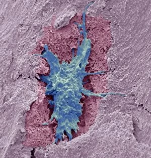
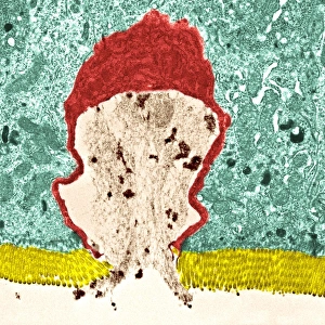
"Unlocking the Secrets: From Osteocytes to Trojan Horses" Delving into the microscopic world, SEM C016 / 9025 reveals the intricate structure of an osteocyte bone cell


All Professionally Made to Order for Quick Shipping
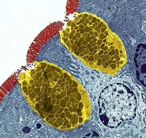
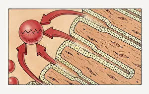
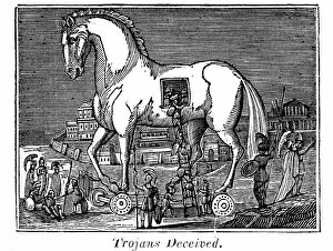
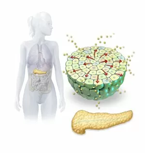
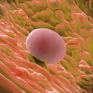
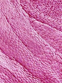
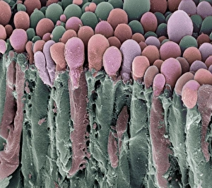
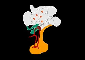
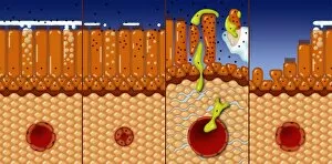
"Unlocking the Secrets: From Osteocytes to Trojan Horses" Delving into the microscopic world, SEM C016 / 9025 reveals the intricate structure of an osteocyte bone cell, hinting at its hidden functions within our skeletal system. Islets of Langerhans cells come alive in a captivating artwork, concealing their vital role in regulating blood sugar levels and secreting hormones like insulin. Another glimpse through SEM C016 / 9026 showcases the mysterious osteocyte bone cell once again, leaving us wondering about its undisclosed contributions to maintaining healthy bones. Gastric glands take center stage in an illustration as they secretly secrete pepsin, an enzyme responsible for breaking down proteins into digestible peptides within our stomachs. Inspired by George Cruikshank's work on Netley Abbey: A Legend of Hampshire, Picture No. 10862034 alludes to hidden tales and concealed histories waiting to be unraveled. The legendary Trojan Horse serves as a metaphor for secrecy as Greek raiding parties cunningly secreted themselves inside this great wooden horse during ancient times. Pavlov's groundbreaking findings on conditioned salivary reflexes are depicted in a diagram that hints at how our organs of touch and temperature play a covert role in triggering certain responses. Goblet cells remain shrouded in mystery until observed under TEM (Transmission Electron Microscope), revealing their unique shape and function in producing mucus throughout our body's various systems. Light micrograph C016 / 0522 unveils the enigmatic landscape of the large intestine, inviting us to explore its hidden intricacies and understand its crucial role in digestion and waste elimination. Artwork showcasing salivary gland anatomy leaves us pondering over these unsung heroes that silently secrete saliva necessary for lubrication during eating and speaking processes.