
Stereocilium Collection
"Stereocilia: Unveiling the Intricate Inner Ear Hairs through SEM Imagery" Delving into the depths of our auditory system


All Professionally Made to Order for Quick Shipping
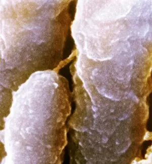
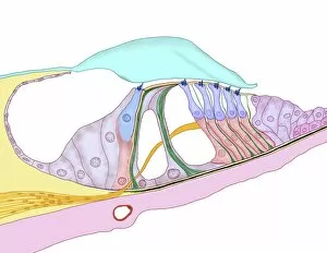
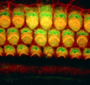
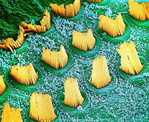
"Stereocilia: Unveiling the Intricate Inner Ear Hairs through SEM Imagery" Delving into the depths of our auditory system, we encounter the mesmerizing world of stereocilia. These delicate inner ear hairs play a vital role in our ability to perceive sound and maintain balance. Through scanning electron microscopy (SEM), their intricate structures are brought to life, revealing a fascinating microcosm within. One captivating artwork showcases the ampullary cupula, an essential component of our vestibular system responsible for detecting rotational movements. Its graceful curves and fine details highlight its crucial function in maintaining equilibrium. Another artwork presents us with a glimpse into the structure of the cochlea, resembling an awe-inspiring spiral staircase. This masterpiece illustrates how these coiled chambers transform sound vibrations into electrical signals that can be interpreted by our brain. Zooming in further with SEM imagery, we witness stunning close-ups of inner ear hair cells - C014/4845, C014/4852, C014/4851, and C014/4844. These microscopic wonders possess bundles of stereocilia protruding from their surfaces like tiny antennae eagerly awaiting sensory input. The ampullary cupula reappears as another artwork capturing its ethereal beauty once more. It reminds us that this structure houses sensory hair cells responsible for detecting changes in head position or movement – crucial information for maintaining balance and spatial orientation. SEM unveils yet another enchanting view – sensory hair cells within our ears' labyrinthine recesses. Their slender stereocilia stand tall like sentinels guarding against silence while enabling us to experience the symphony of sounds around us. These inner ear sensory hairs serve as nature's own orchestra conductor; they convert mechanical vibrations into electrical signals that ignite sensations within us—allowing melodies to dance upon eardrums and harmonies to resonate deep within our souls.