
Transmission Electron Microgra Collection
"Exploring the Microscopic World: Unveiling Intricate Structures through Transmission Electron Micrography" In the realm of cellular biology


All Professionally Made to Order for Quick Shipping
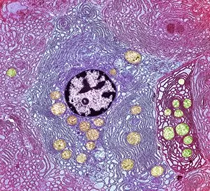
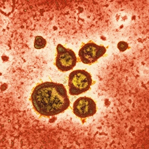
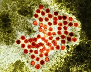
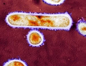
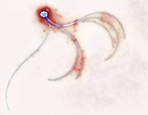
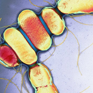
"Exploring the Microscopic World: Unveiling Intricate Structures through Transmission Electron Micrography" In the realm of cellular biology, transmission electron micrography (TEM) has revolutionized our understanding of various organisms and their intricate structures. This cutting-edge technique allows us to delve into the hidden world of cells and viruses, revealing fascinating details that were once invisible to the naked eye. Peering deep within a pancreas cell using TEM, we witness its complex machinery at work, unraveling the secrets behind insulin production and regulation. Moving on to the small intestine, TEM exposes its densely packed villi and microvilli, responsible for nutrient absorption. Zooming further down into viral dimensions, TEM captures norovirus particles in all their glory – tiny spheres with characteristic spikes that cause gastrointestinal distress. Parvovirus particles also come into focus; these minute entities are known for causing diseases in animals but can occasionally infect humans as well. Shifting gears towards hepatitis A virus particles observed via TEM reveals their distinctive shape resembling miniature soccer balls – an insight crucial for developing effective vaccines against this liver-infecting pathogen. Meanwhile, influenza viruses take center stage as both Influenzavirus B and Influenzavirus A showcase their unique morphologies under TEM's watchful lens. Delving deeper into avian influenza virus particles unveils peculiar elongated shapes reminiscent of rods or filaments - a stark contrast to other flu strains captured by TEM before. Returning again to Influenzavirus B reminds us of its spherical structure adorned with surface projections called hemagglutinin spikes. Lastly, astrovirus particles make an appearance through TEM imagery - showcasing star-like formations that give them their name while being associated with gastroenteritis in humans. Transmission electron micrography continues to be instrumental in unraveling nature's microscopic wonders.