Fraud Blocker Skip to main content

Tunica Intima Collection

The tunica intima

Background imageTunica Intima Collection: Heart with arteries showing cholesterol in one and plaque in the other

Heart with arteries showing cholesterol in one and plaque in the other
Heart with arteries showing cholesterol in one artery and atherosclerotic plaque in the other

Background imageTunica Intima Collection: Arteries on heart showing atherosclerotic plaque in an artery

Arteries on heart showing atherosclerotic plaque in an artery

Background imageTunica Intima Collection: Build-up of atherosclerotic plaque in an artery

Build-up of atherosclerotic plaque in an artery

Background imageTunica Intima Collection: Acute coronary syndrome - microvascular obstruction

Acute coronary syndrome - microvascular obstruction

Background imageTunica Intima Collection: Interior view of heart with detail of muscle cells and atherosclerotic artery

Interior view of heart with detail of muscle cells and atherosclerotic artery. Chest x-ray in background

Background imageTunica Intima Collection: Thrombus forming on valve within vein

Thrombus forming on valve within vein

Background imageTunica Intima Collection: Normal artery compared to plaque and thrombus formation in artery

Normal artery compared to plaque and thrombus formation in artery

Background imageTunica Intima Collection: Artery showing atherosclerotic plaque, platelets and red blood cells

Artery showing atherosclerotic plaque, platelets and red blood cells
Close-up of artery showing atherosclerotic plaque, platelets and red blood cells

Background imageTunica Intima Collection: Blood vessel anatomy

Blood vessel anatomy

Background imageTunica Intima Collection: Anatomy of human veins and arteries, black background

Anatomy of human veins and arteries, black background

Background imageTunica Intima Collection: Human arteries, artwork F008 / 1037

Human arteries, artwork F008 / 1037
Human arteries, computer artwork

Background imageTunica Intima Collection: Human arteries, artwork F008 / 1025

Human arteries, artwork F008 / 1025
Human arteries, computer artwork

Background imageTunica Intima Collection: Human arteries, artwork F008 / 1016

Human arteries, artwork F008 / 1016
Human arteries, computer artwork

Background imageTunica Intima Collection: Female cardiovascular system, artwork F008 / 1009

Female cardiovascular system, artwork F008 / 1009
Female cardiovascular system, computer artwork

Background imageTunica Intima Collection: Human arteries, artwork F008 / 1024

Human arteries, artwork F008 / 1024
Human arteries, computer artwork

Background imageTunica Intima Collection: Female cardiovascular system, artwork F008 / 1010

Female cardiovascular system, artwork F008 / 1010
Female cardiovascular system, computer artwork

Background imageTunica Intima Collection: Human arteries, artwork F008 / 1036

Human arteries, artwork F008 / 1036
Human arteries, computer artwork

Background imageTunica Intima Collection: Human arteries, artwork F008 / 1023

Human arteries, artwork F008 / 1023
Human arteries, computer artwork

Background imageTunica Intima Collection: Coloured SEM of section through a human arteriole

Coloured SEM of section through a human arteriole
Arteriole. Coloured scanning electron micrograph (SEM) of a cross-section through a small human artery known as an arteriole

Background imageTunica Intima Collection: Contracted artery, TEM

Contracted artery, TEM
Contracted elastic artery. Coloured transmission electron micrograph (TEM) of a section through an artery. Red blood cells (erythrocytes) are seen in the lumen (top centre)

Background imageTunica Intima Collection: Artery anatomy, artwork

Artery anatomy, artwork
Artery anatomy, computer artwork. At the centre of the artery is the lumen, with three red blood cells (erythrocytes, bottom right)

Background imageTunica Intima Collection: Arteriole, SEM

Arteriole, SEM
Arteriole. Coloured scanning electron micrograph (SEM) of a cross-section through a small human artery known as an arteriole. Red blood cells and some fibrin fibres are seen in the central lumen

Background imageTunica Intima Collection: Arteriole and red blood cells, SEM

Arteriole and red blood cells, SEM
Arteriole. Coloured scanning electron micrograph (SEM) of a cross-section through a small, thin- walled artery known as an arteriole

Background imageTunica Intima Collection: Muscular artery

Muscular artery. Light micrograph of a transverse section through a muscular artery. This blood vessel has a thick, muscular wall that enables it to carry blood under pressure

Background imageTunica Intima Collection: Artery, TEM

Artery, TEM
Artery. Coloured transmission electron micrograph (TEM) of a transverse section through an artery. Red blood cells (erythrocytes, red) and white blood cells (leucocytes)

Background imageTunica Intima Collection: Blood vessel structure, artwork

Blood vessel structure, artwork
Blood vessel structure. Artwork showing the layers of tissue in the walls of a vein. In the lumen of the vein are valves (grey) that prevent the back flow of blood

Background imageTunica Intima Collection: Heart blood vessel wall, TEM

Heart blood vessel wall, TEM


All Professionally Made to Order for Quick Shipping

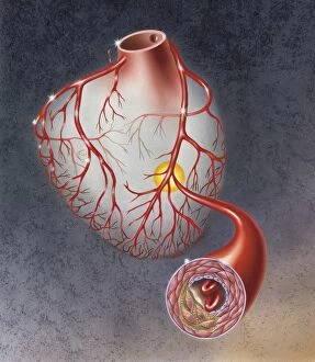
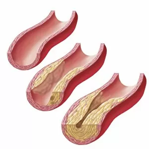
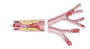
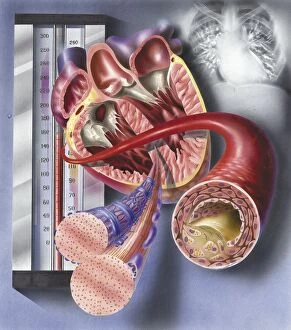
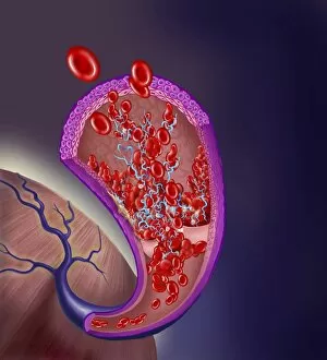
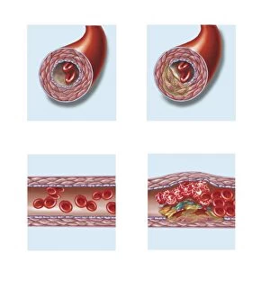
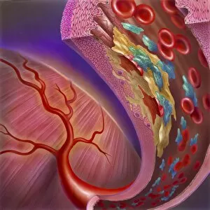
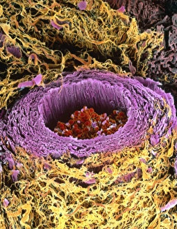
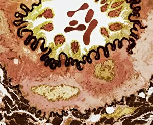
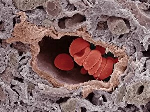
The tunica intima: Exploring the intricate world within our blood vessels A closer look at the heart reveals a stark contrast between two arteries - one showcasing cholesterol build-up, while the other displays atherosclerotic plaque. Witnessing an artery affected by atherosclerosis, we realize the severity of plaque formation and its impact on cardiovascular health. The gradual accumulation of atherosclerotic plaque in an artery serves as a reminder of the importance of maintaining healthy blood vessels. Microvascular obstruction caused by acute coronary syndrome unveils how even tiny obstructions can have significant consequences for heart health. Peering into the interior view of the heart, we observe muscle cells alongside an atherosclerotic artery, emphasizing the need to protect our cardiovascular system from potential damage. Within veins, thrombus formation on valves poses risks that should not be ignored – highlighting why venous health is just as crucial as arterial well-being. Comparing normal arteries with those affected by plaque and thrombus formation underscores how vital it is to prevent these detrimental changes within our circulatory system. An image capturing an artery exhibiting both atherosclerotic plaque, platelets, and red blood cells reminds us that maintaining vascular health requires ongoing attention and care. Conceptually exploring blood vessels allows us to appreciate their complexity and understand why safeguarding their integrity is paramount for overall well-being.