Home > Popular Themes > Human Body
Contracted artery, TEM

![]()
Wall Art and Photo Gifts from Science Photo Library
Contracted artery, TEM
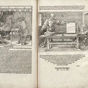
Contracted elastic artery. Coloured transmission electron micrograph (TEM) of a section through an artery. Red blood cells (erythrocytes) are seen in the lumen (top centre). The lumen is lined with a layer of epithelium (yellow with red nuclei) and an inner elastic membrane (black). These together make up the tunica intima. This is surrounded by a layer of smooth muscle (pink) called the tunica media. The layer of muscle has contracted to decrease the volume of the lumen, causing the tunica intima to become folded. The outer layer of the artery, the tunica adventitia (brown), which contains collagen, is seen at bottom. An outer elastic membrane (black) lies at the junction of the tunica media and adventitia
Science Photo Library features Science and Medical images including photos and illustrations
Media ID 6446573
© STEVE GSCHMEISSNER/SCIENCE PHOTO LIBRARY
Blood Collagen Contracted Contracting Cross Section Endothelium Erythrocyte Erythrocytes Fibres Lumen Red Blood Cell Red Blood Cells Smooth Muscle Transmission Electron Micrograph Transmission Electron Microscope Tunica Adventitia Tunica Intima Tunica Media Vessel Vessels Artery False Coloured Section Sectioned
EDITORS COMMENTS
This print from Science Photo Library showcases a contracted elastic artery in all its intricate glory. Through the lens of a transmission electron microscope (TEM), we are granted a mesmerizing glimpse into the inner workings of our circulatory system. At the center, red blood cells elegantly float within the lumen, surrounded by a layer of yellow epithelium adorned with red nuclei. Just beneath lies an inner elastic membrane, painted black and forming part of the tunica intima. This delicate structure is encased by a pink layer known as the tunica media – composed of smooth muscle that has skillfully contracted to reduce the lumen's volume. As a result, folds appear on the surface of the tunica intima. The bottom portion reveals another crucial component: The tunica adventitia, represented in rich brown hues and containing collagen fibers that provide strength and support to this vital vessel. Separating it from the tunica media is an outer elastic membrane, adding yet another layer of complexity to this microscopic marvel. This false-colored TEM image not only highlights biological intricacies but also invites contemplation about our own bodies' remarkable capabilities. It serves as a reminder that even at such minuscule scales, beauty can be found in every corner – reminding us how truly awe-inspiring nature can be.
MADE IN AUSTRALIA
Safe Shipping with 30 Day Money Back Guarantee
FREE PERSONALISATION*
We are proud to offer a range of customisation features including Personalised Captions, Color Filters and Picture Zoom Tools
SECURE PAYMENTS
We happily accept a wide range of payment options so you can pay for the things you need in the way that is most convenient for you
* Options may vary by product and licensing agreement. Zoomed Pictures can be adjusted in the Cart.