Fraud Blocker Skip to main content

Uterine Collection

"Exploring the Intricacies of the Uterine Journey

Background imageUterine Collection: Uterus lining during menstruation, SEM

Uterus lining during menstruation, SEM
Uterus during menstruation. Coloured scanning electron micrograph (SEM) of the lining of the uterus being shed during menstruation

Background imageUterine Collection: IUD contraceptive and sperm cells

IUD contraceptive and sperm cells. Computer artwork of an intrauterine device (IUD, red and white, centre) in a womans uterus, surrounded by sperm cells (white, not to scale)

Background imageUterine Collection: Birth of two stillborn babies, 15th century (engraving)

Birth of two stillborn babies, 15th century (engraving)
5311584 Birth of two stillborn babies, 15th century (engraving); (add.info.: The explosion is supposed to be uterine gasses)

Background imageUterine Collection: Albinus, Uterus II, Pl. V, illustration from Tabulae ossium humanorum

Albinus, Uterus II, Pl. V, illustration from Tabulae ossium humanorum
HMB306116 Albinus, Uterus II, Pl. V, illustration from Tabulae ossium humanorum, by Bernhard Siegfried Albinus (1697-1770), published by J.&H. Verbeek, bibliop

Background imageUterine Collection: Albinus, Uterus, Pl. III, illustration from Tabulae ossium humanorum

Albinus, Uterus, Pl. III, illustration from Tabulae ossium humanorum
HMB306115 Albinus, Uterus, Pl. III, illustration from Tabulae ossium humanorum, by Bernhard Siegfried Albinus (1697-1770), published by J.&H. Verbeek, bibliop

Background imageUterine Collection: Albinus, Uterus, Pl. II, illustration from Tabulae ossium humanorum

Albinus, Uterus, Pl. II, illustration from Tabulae ossium humanorum
HMB306114 Albinus, Uterus, Pl. II, illustration from Tabulae ossium humanorum, by Bernhard Siegfried Albinus (1697-1770), published by J.&H. Verbeek, bibliop

Background imageUterine Collection: 18th century tweezers and speculum for obstetrics

18th century tweezers and speculum for obstetrics
Wire-knot tweezers for obstetrics 1, 2, use of the wire-knot tweezer to remove an uterine polyp 3, tweezers and speculum invented by Andre Levret 4, 5

Background imageUterine Collection: Uterus, endoscope view C017 / 6805

Uterus, endoscope view C017 / 6805
Uterus. Endoscope (laparascope) view of a healthy uterus (womb, large, upper centre), with the two fallopian tubes (left and right) and an ovary (white, lower right)

Background imageUterine Collection: Uterine fibroids, MRI scan C018 / 0466

Uterine fibroids, MRI scan C018 / 0466
Uterine fibroids. Magnetic resonance imaging (MRI) scan of the pelvis of a 47 year old female with two large fibroids (centre) in her uterus

Background imageUterine Collection: Uterine fibroid C015 / 6407

Uterine fibroid C015 / 6407
Uterine fibroid. Gross specimen of a sectioned uterine fibroid (left). A fibroid is a fibrous benign tumour originating from muscular tissue

Background imageUterine Collection: Endometrial polyp, ultrasound scan C017 / 7800

Endometrial polyp, ultrasound scan C017 / 7800
Endometrial polyp. Ultrasound scan of the uterus (womb) of a 42 year old female patient with a polyp (lower centre) in the endometrium, the lining of the womb

Background imageUterine Collection: Heavy menstrual bleeding, artwork

Heavy menstrual bleeding, artwork
Heavy menstrual bleeding, conceptual computer artwork. Collection of measuring cylinders containing differing amounts of blood

Background imageUterine Collection: John Towneley

John Towneley
JOHN TOWNELEY of Towneley, uterine brother (same mother, different father) of Alexander Nowell Date: 1528? - 1607

Background imageUterine Collection: Fimbriae of a fallopian tube, SEM C016 / 9047

Fimbriae of a fallopian tube, SEM C016 / 9047
Fimbriae of a fallopian tube, coloured scanning electron micrograph (SEM). Fimbriae are the folds at the opening (ostium) of a fallopian tube, next to an ovary

Background imageUterine Collection: Fimbriae of a fallopian tube, SEM C016 / 9048

Fimbriae of a fallopian tube, SEM C016 / 9048
Fimbriae of a fallopian tube, coloured scanning electron micrograph (SEM). Fimbriae are the folds at the opening (ostium) of a fallopian tube, next to an ovary

Background imageUterine Collection: Fimbriae of a fallopian tube, SEM C016 / 9049

Fimbriae of a fallopian tube, SEM C016 / 9049
Fimbriae of a fallopian tube, coloured scanning electron micrograph (SEM). Fimbriae are the folds at the opening (ostium) of a fallopian tube, next to an ovary

Background imageUterine Collection: Fimbriae of a fallopian tube, SEM C016 / 9046

Fimbriae of a fallopian tube, SEM C016 / 9046
Fimbriae of a fallopian tube, coloured scanning electron micrograph (SEM). Fimbriae are the folds at the opening (ostium) of a fallopian tube, next to an ovary

Background imageUterine Collection: Uterine fibroids, MRI scan C018 / 0467

Uterine fibroids, MRI scan C018 / 0467
Uterine fibroids. Coloured magnetic resonance imaging (MRI) scan of the pelvis of a 47 year old female with two large fibroids (pink) in her uterus

Background imageUterine Collection: Uterine fibroids, MRI scan C018 / 0468

Uterine fibroids, MRI scan C018 / 0468
Uterine fibroids. Coloured magnetic resonance imaging (MRI) scan of the pelvis of a 47 year old female with two large fibroids (pink) in her uterus

Background imageUterine Collection: Uterine fibroids, MRI scan C018 / 0469

Uterine fibroids, MRI scan C018 / 0469
Uterine fibroids. Coloured magnetic resonance imaging (MRI) scan of the pelvis of a 47 year old female with two large fibroids (pink) in her uterus

Background imageUterine Collection: Uterine fibroid, X-ray C014 / 4919

Uterine fibroid, X-ray C014 / 4919
Uterus tumour. Coloured X-ray of the abdomen of a woman with a fibroid (round, centre) in her uterus. A fibroid is a fibrous benign tumour originating from muscular tissue (leimyoma)

Background imageUterine Collection: Uterine fibroid, X-ray C014 / 4918

Uterine fibroid, X-ray C014 / 4918
Uterus tumour. Coloured X-ray of the abdomen of a woman with a fibroid (orange, round) in her uterus. A fibroid is a fibrous benign tumour originating from muscular tissue (leimyoma)

Background imageUterine Collection: Endometriosis, X-ray C014 / 4917

Endometriosis, X-ray C014 / 4917
Endometriosis. Coloured X-ray of the abdomen of woman with endometriosis. Endometriosis is the movement of cells from the lining of the uterus to the abdominal cavity

Background imageUterine Collection: Endometriosis, X-ray C014 / 4916

Endometriosis, X-ray C014 / 4916
Endometriosis. Coloured X-ray of the abdomen of woman with endometriosis. Endometriosis is the movement of cells from the lining of the uterus to the abdominal cavity

Background imageUterine Collection: Uterine arteries, 1825 artwork

Uterine arteries, 1825 artwork
Uterine arteries. Dissection showing the arteries (red) of the uterus (lower centre) of a woman who died six days after giving birth. The ovaries and fallopian tubes are either side

Background imageUterine Collection: Endometrial hyperplasia, light micrograph C015 / 7104

Endometrial hyperplasia, light micrograph C015 / 7104
Endometrial hyperplasia. Light micrograph of a section through tissue from the endometrium, in a case of endometrial hyperplasia. The endometrium is the uterus lining

Background imageUterine Collection: Uterine fibroid, light micrograph C015 / 7101

Uterine fibroid, light micrograph C015 / 7101
Uterine fibroid. Light micrograph of a section through a uterine fibroid (centre). A fibroid is a fibrous benign tumour originating from muscular tissue

Background imageUterine Collection: Cervical smear showing endometrial cancer C015 / 6750

Cervical smear showing endometrial cancer C015 / 6750
Cervical smear showing endometrial cancer. Light micrograph of cells from a cervical smear test showing endometrial sarcoma. This is a cancer of the lining of the womb, the endometrium

Background imageUterine Collection: Uterine fibroid, light micrograph C015 / 6737

Uterine fibroid, light micrograph C015 / 6737
Uterine fibroid. Light micrograph of a section through a uterine fibroid (oval). A fibroid is a fibrous benign tumour originating from muscular tissue

Background imageUterine Collection: Secondary endometrial cancer C015 / 6058

Secondary endometrial cancer C015 / 6058
Secondary endometrial cancer. Light micrograph of a section through a tumour in an inguinal lymph node due to a secondary endometrial cancer

Background imageUterine Collection: Uterine fibroid C015 / 6416

Uterine fibroid C015 / 6416
Uterine fibroid. Gross specimen of a sectioned uterine fibroid. A fibroid is a fibrous benign tumour originating from muscular tissue

Background imageUterine Collection: Uterine fibroid, light micrograph C015 / 6413

Uterine fibroid, light micrograph C015 / 6413
Uterine fibroid. Light micrograph of a section through a uterine fibroid. A fibroid is a fibrous benign tumour originating from muscular tissue

Background imageUterine Collection: Uterine fibroid C015 / 6415

Uterine fibroid C015 / 6415
Uterine fibroid. Gross specimen of a sectioned uterine fibroid. A fibroid is a fibrous benign tumour originating from muscular tissue

Background imageUterine Collection: Uterine fibroid, light micrograph C015 / 6411

Uterine fibroid, light micrograph C015 / 6411
Uterine fibroid. Light micrograph of a section through a uterine fibroid (centre). A fibroid is a fibrous benign tumour originating from muscular tissue

Background imageUterine Collection: Uterine fibroid, light micrograph C015 / 6412

Uterine fibroid, light micrograph C015 / 6412
Uterine fibroid. Light micrograph of a section through a uterine fibroid (oval). A fibroid is a fibrous benign tumour originating from muscular tissue

Background imageUterine Collection: Uterine fibroid C015 / 6406

Uterine fibroid C015 / 6406
Uterine fibroid. Gross specimen of a uterine fibroid. A fibroid is a fibrous benign tumour originating from muscular tissue

Background imageUterine Collection: Endometrial polyp, ultrasound scan C017 / 7799

Endometrial polyp, ultrasound scan C017 / 7799
Endometrial polyp. Doppler ultrasound scan of the uterus (womb) of a 42 year old female patient with a polyp (lower centre) in the endometrium, the lining of the womb

Background imageUterine Collection: Internal wall of uterus, SEM

Internal wall of uterus, SEM
Internal wall of uterus. Coloured scanning electron micrograph of the mucosa, also known as endometrium, lining the body of the human uterus

Background imageUterine Collection: Uterine lining, SEM

Uterine lining, SEM
Uterine lining. Coloured scanning electron micrograph (SEM) of the lining of a uterus. This is the endometrium, a highly-folded mucous membrane that is covered with ciliated epithelial cells

Background imageUterine Collection: Blocked Fallopian tubes, X-ray

Blocked Fallopian tubes, X-ray
Blocked Fallopian tubes. Coloured X-ray of a womans reproductive system, with her uterus (green) and blocked Fallopian tubes (oviducts, yellow)

Background imageUterine Collection: Heavy menstrual bleeding

Heavy menstrual bleeding. Conceptual computer artwork of red blood cells pouring out a tap, representing the heavy bleeding seen in menorrhagia, or excessive menstrual bleeding

Background imageUterine Collection: Uterine fibroid, MRI scan

Uterine fibroid, MRI scan

Background imageUterine Collection: Uterus cells, TEM

Uterus cells, TEM
Uterus cells. Coloured transmission electron micrograph (TEM) of endometrial cells (yellow) from the uterus. These cells form the endometrium, the epithelial tissue lining inside the uterus


All Professionally Made to Order for Quick Shipping

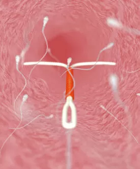
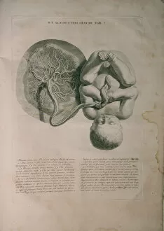
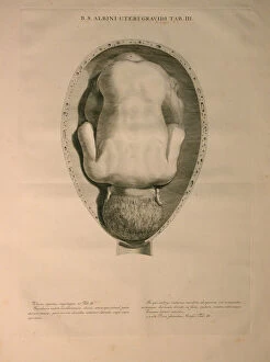
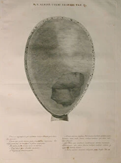
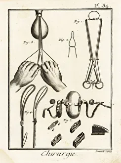
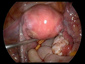
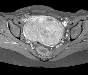
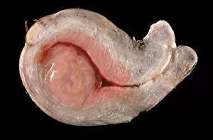
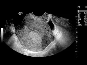
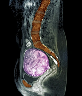
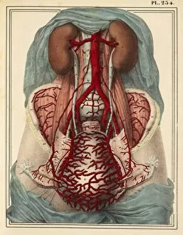
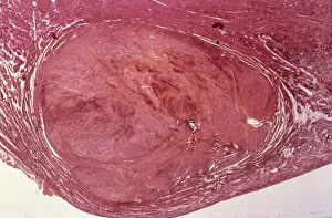
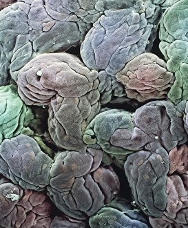
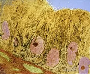
"Exploring the Intricacies of the Uterine Journey: From Menstruation to Childbirth" The fascinating process of menstruation unveils the intricate dance between hormones and the uterus lining, as it sheds during this natural cycle. SEM images capture this mesmerizing phenomenon. In the realm of contraception, IUDs stand tall as a reliable choice. These tiny devices work by preventing sperm cells from reaching their destination in the uterine cavity. Witnessing childbirth is witnessing pure magic unfold within the uterine walls. As labor progresses, uterine cells undergo remarkable changes to accommodate new life entering into this world. Albinus's detailed illustrations from Tabulae ossium humanorum offer a glimpse into our understanding anatomy throughout history – Pl. V showcases its complexity and beauty. Continuing with Albinus's work, Pl. III takes us on a visual journey through different layers and structures within the uterus, shedding light on its intricacies like never before. Delving deeper into anatomical exploration, Pl. II reveals even more hidden wonders within the uterus – an organ that has captivated scientists and artists alike for centuries. Obstetrics tools have come a long way since John Towneley's 18th-century tweezers and speculum were used for delicate procedures related to childbirth – reminding us of how far medical advancements have taken us today. With modern technology like endoscopes providing unprecedented views inside our bodies, we can now marvel at live footage capturing every detail of the uterus in motion (C017 / 6805). Uterine fibroids are common growths that occur in many women's reproductive years; MRI scans (C018 / 0466) help doctors diagnose these benign tumors accurately while ensuring appropriate treatment plans are put in place. Heavy menstrual bleeding can be debilitating for some individuals, impacting their daily lives.