Home > Popular Themes > Human Body
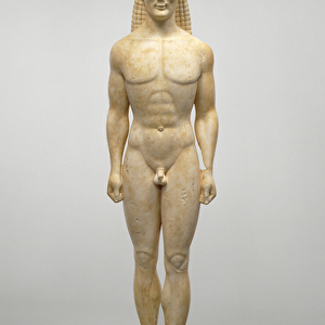
Uterine arteries, 1825 artwork

![]()
Wall Art and Photo Gifts from Science Photo Library
Uterine arteries, 1825 artwork
Uterine arteries. Dissection showing the arteries (red) of the uterus (lower centre) of a woman who died six days after giving birth. The ovaries and fallopian tubes are either side. Within the abdominal cavity are muscles (orange), the aorta and iliac arteries (red), kidneys (brown) and ureters (white). This anatomical artwork is plate 234 from volume 4 of Manuel d anatomie descriptive du corps humain (1825). This 5-volume anatomy atlas was produced by French physician and surgeon Jules Germain Cloquet (1790-1883). The illustrations were by Haincelin. Volume 4 illustrated the anatomy of the circulatory and respiratory systems
Science Photo Library features Science and Medical images including photos and illustrations
Media ID 9222895
© SCIENCE PHOTO LIBRARY
1825 Abdomen Abdominal Abdominal Cavity Anatomical Artwork Anatomical Illustration Anatomy Atlas Anterior Aorta Arterial System Arteries Blood Vessels Childbirth Death Dissected Dissection Fallopian Tube Female Reproductive System French Frontal Gynaecology Gynecology Haincelin Iliac Artery Jules Germain Cloquet Kidneys Muscles Obstetrics Organs Ovaries Ovary Oxygenated Blood Reproductive System Tubes Ureters Uterine Uterus Vascular Volume 4 Volume Iv Womb Artery Blood Vessel Circulatory System
EDITORS COMMENTS
This 19th-century artwork, titled "Uterine Arteries" offers a glimpse into the intricate anatomy of the female reproductive system. Created in 1825 by French physician and surgeon Jules Germain Cloquet, this illustration is part of his renowned five-volume anatomy atlas. In this particular print, we see a dissection of the uterus (lower center) from a woman who tragically passed away just six days after giving birth. The ovaries and fallopian tubes flank either side, while surrounding organs such as muscles, kidneys, and ureters are depicted within the abdominal cavity. The vibrant red color highlights the uterine arteries coursing through the illustration, symbolizing their vital role in supplying oxygenated blood to support pregnancy. This detailed anatomical representation provides valuable insights into gynecology and obstetrics during that era. Crafted with precision by artist Haincelin under Cloquet's guidance, this artwork showcases both scientific accuracy and artistic finesse. It serves as a testament to medical advancements in understanding the circulatory system's role in reproduction. As we gaze upon this historical masterpiece today, it reminds us of how far our knowledge has progressed since its creation. Yet it also pays homage to those early pioneers like Jules Germain Cloquet who dedicated their lives to unraveling the mysteries of human anatomy for generations to come.
MADE IN AUSTRALIA
Safe Shipping with 30 Day Money Back Guarantee
FREE PERSONALISATION*
We are proud to offer a range of customisation features including Personalised Captions, Color Filters and Picture Zoom Tools
SECURE PAYMENTS
We happily accept a wide range of payment options so you can pay for the things you need in the way that is most convenient for you
* Options may vary by product and licensing agreement. Zoomed Pictures can be adjusted in the Cart.