
White Blood Cell Collection (page 8)
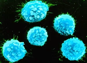
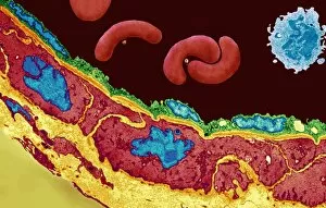
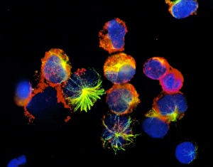
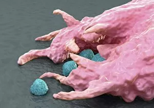
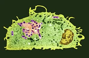
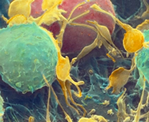
"Guardians of Health: Exploring the Mighty White Blood Cell" T lymphocytes and cancer cells: Unveiling the battle within, as T lymphocytes wage war against cancer cells


All Professionally Made to Order for Quick Shipping
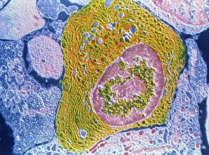
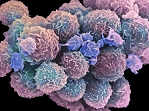
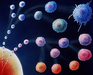
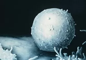
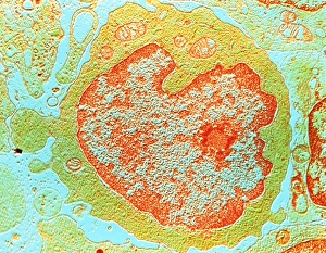
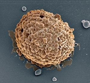
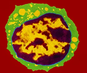
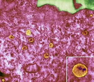
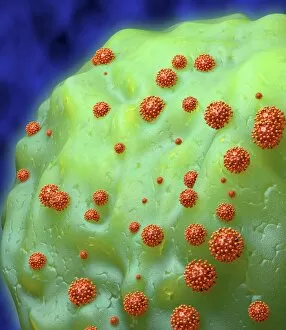
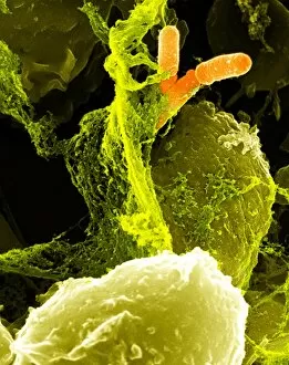
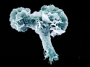
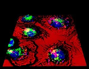
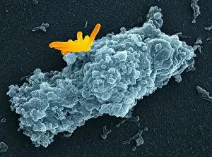
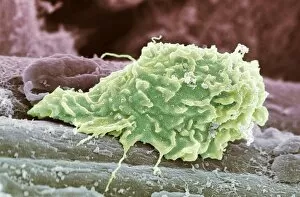
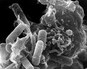
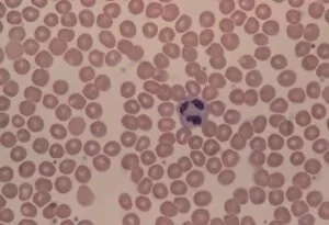
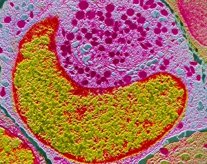
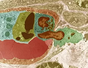
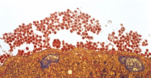
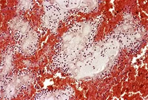
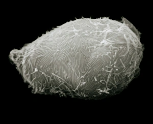
"Guardians of Health: Exploring the Mighty White Blood Cell" T lymphocytes and cancer cells: Unveiling the battle within, as T lymphocytes wage war against cancer cells, SEM C001 / 1679. Neutrophil engulfing MRSA: Witness the incredible defense mechanism as a neutrophil engulfs MRSA bacteria, SEM C018 / 8596. Dendritic cells artwork: Unlocking the secrets of immune response with stunning artwork depicting dendritic cells at work. TEM of human white blood cell bearing HLA antigen: Peering into the intricate world of immunity through a transmission electron microscope image showcasing a white blood cell displaying HLA antigens. Blood cells in harmony: A mesmerizing glimpse into our life force - an ensemble of diverse blood cells working together for our well-being. Coloured SEM of a white blood cell (lymphocyte): Dive deep into the vibrant realm of lymphocytes captured in this captivating colored scanning electron microscope image. Basophil white blood cell: Discovering the lesser-known heroes among us - basophilic white blood cells that play crucial roles in allergic reactions and inflammation. Bacteria infecting a macrophage, SEM: Witness how macrophages confront invading bacteria head-on under high-resolution scanning electron microscopy imagery. Blood coagulation cascade artwork: Unraveling the intricacies behind clot formation with an artistic representation capturing every step in this vital process, artwork C016 / 9873 Red and white blood cells, SEM : Marvel at nature's palette as red and white blood cells come alive under scanning electron microscopy - an exquisite symphony within us all. Blood clot, SEM C016 / 9747 : Delving into hemostasis marvels with a striking scanning electron micrograph revealing intricate details within a formed clot structure. Dohle bodies in blood cell, micrograph.