Fraud Blocker Skip to main content

White Blood Cell Collection (page 4)

"Guardians of Health: Exploring the Mighty White Blood Cell" T lymphocytes and cancer cells: Unveiling the battle within, as T lymphocytes wage war against cancer cells

Background imageWhite Blood Cell Collection: Activated plasma cell, TEM

Activated plasma cell, TEM
Activated plasma cell, coloured transmission electron micrograph (TEM). Plasma cells are derived from stimulated B lymphocytes (white blood cells)

Background imageWhite Blood Cell Collection: Basophil white blood cell, TEM

Basophil white blood cell, TEM
Basophil white blood cell. Transmission electron micrograph (TEM) of section through a circulating basophil cell. Basophils are the smallest and least common of the white blood cells

Background imageWhite Blood Cell Collection: Lymphoblastic sarcoma, light micrograph

Lymphoblastic sarcoma, light micrograph
Lymphoblastic sarcoma. Light micrograph of blood cells from bone marrow in a case of lymphoblastic sarcoma. These small lymphoblasts (dark red) characteristically have conspicuous nucleoli

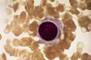
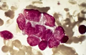
Background imageWhite Blood Cell Collection: Plasmocyte blood cell, light micrograph

Plasmocyte blood cell, light micrograph. Plasmocytes, also called plasma cells, are derived from stimulated B lymphocytes (white blood cells)
Background imageWhite Blood Cell Collection: Multiple myeloma, light micrograph

Multiple myeloma, light micrograph
Multiple myeloma. Light micrograph of dystrophy (abnormality) of plasmocyte blood cells from bone marrow in a case of multiple myeloma, also known as Kahlers disease or plasma cell myeloma
Background imageWhite Blood Cell Collection: Lymphoplasmacytic lymphoma, micrograph

Lymphoplasmacytic lymphoma, micrograph
Lymphoplasmacytic lymphoma. Light micrograph of a mixture of small lymphocytes and plasma cells from bone marrow in a case of lymphoplasmacytic lymphoma, also known as Waldenstroms macroglobulinemia

Background imageWhite Blood Cell Collection: Hairy cell leukaemia, light micrograph

Hairy cell leukaemia, light micrograph
Hairy cell leukaemia. Light micrograph of blood cells in a case of hairy cell leukaemia. This sample, treated with May-Grunwald-Giemsa (MGG) stain

Background imageWhite Blood Cell Collection: Mast cells, light micrograph

Mast cells, light micrograph
Mast cells. Light micrograph of several mast cells (pink rounded objects). These leucocytes (white blood cells) contain the chemical mediators histamine, serotonin and heparin

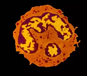
Background imageWhite Blood Cell Collection: Lymphocyte white blood cell, TEM

Lymphocyte white blood cell, TEM
Lymphocyte white blood cell. Transmission electron micrograph (TEM) of a section through lymphocyte, showing the nucleus (large, round, centre), endoplasmic reticulum (ER)

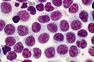
Background imageWhite Blood Cell Collection: Acute lymphoblastic leukaemia, micrograph

Acute lymphoblastic leukaemia, micrograph
Acute lymphoblastic leukaemia. Light micrograph of blood cells from bone marrow in a case of acute lymphoblastic leukaemia. Leukaemia is a cancer where certain blood cells form in excess

Background imageWhite Blood Cell Collection: Malignant histiocytosis, light micrograph

Malignant histiocytosis, light micrograph
Malignant histiocytosis. Light micrograph of blood cells in a case of malignant histiocytosis. Hystiocytosis is an excess number of histiocytes

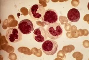
Background imageWhite Blood Cell Collection: Blood cells, light micrograph

Blood cells, light micrograph
Blood cells. Light micrograph of red blood cells (erythrocytes, light blue) and white blood cells (leucocytes, nuclei stained purple)

Background imageWhite Blood Cell Collection: Chediak-Higashi syndrome, micrograph

Chediak-Higashi syndrome, micrograph
Chediak-Higashi syndrome. Light micrograph of blood cells in a case of Chediak-Higashi syndrome. The cells include neutrophil white blood cells with abnormal granules

Background imageWhite Blood Cell Collection: Metastatic breast cancer, micrograph

Metastatic breast cancer, micrograph
Metastatic breast cancer. Light micrograph of blood cells in a case of metastatic (secondary) medullary breast cancer. Medullary breast cancer is a rare form of breast cancer that is diagnosed by

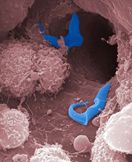
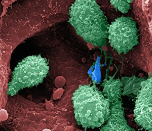
Background imageWhite Blood Cell Collection: Sleeping sickness parasites, SEM

Sleeping sickness parasites, SEM
Sleeping sickness parasites. Coloured scanning electron micrograph (SEM) of a Trypanosoma brucei parasites (blue) and white blood cells (pink) in a mouse liver. T

Background imageWhite Blood Cell Collection: Metastatic lung cancer, micrograph

Metastatic lung cancer, micrograph
Metastatic lung cancer. Light micrograph of blood cells in a case of metastatic (secondary) medullary lung cancer. Medullary cancer (carcinoma)

Background imageWhite Blood Cell Collection: Alders anomaly, light micrograph

Alders anomaly, light micrograph
Alders anomaly. Light micrograph of an abnormal lymphocyte white blood cell (centre) from a blood sample in a case of Alders anomaly

Background imageWhite Blood Cell Collection: Granulocyte white blood cell, TEM

Granulocyte white blood cell, TEM
Granulocyte white blood cell. Coloured transmission electron micrograph (TEM) of a neutrophil granulocyte, a type of white blood cell

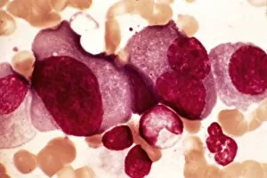
Background imageWhite Blood Cell Collection: Acute leukaemia, light micrograph

Acute leukaemia, light micrograph
Acute leukaemia. Light micrograph of blood cells from bone marrow in a case of acute leukaemia. These cells include a polynuclear degranulated basophil and myeloblasts

Background imageWhite Blood Cell Collection: Sleeping sickness parasite, SEM

Sleeping sickness parasite, SEM
Sleeping sickness parasite. Coloured scanning electron micrograph (SEM) of a Trypanosoma brucei parasite (blue) and a white blood cell (green) in a mouse liver. T

Background imageWhite Blood Cell Collection: Leukaemia cell, SEM

Leukaemia cell, SEM
Leukaemia cell. Coloured scanning electron micrograph (SEM) of a leukaemic (cancerous) lymphocyte white blood cell (green), amongst normal red blood cells (erythrocytes, red)

Background imageWhite Blood Cell Collection: Chronic myeloid leukaemia, micrograph

Chronic myeloid leukaemia, micrograph
Chronic myeloid leukaemia. Light micrograph of blood cells from bone marrow in a case of chronic myeloid leukaemia. Leukaemia is a cancer where certain blood cells form in excess

Background imageWhite Blood Cell Collection: Granulocyte blood cells, light micrograph

Granulocyte blood cells, light micrograph
Granulocyte blood cells. Light micrograph of two types of granulocytes, a type of white blood cell (leucocyte). At left is a neutrophil, with an eosinophil at right

Background imageWhite Blood Cell Collection: Langerhans cell histiocytosis, micrograph

Langerhans cell histiocytosis, micrograph
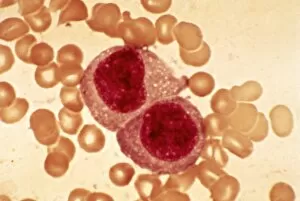
Langerhans cell histiocytosis. Light micrograph of blood cells in a case of Langerhans cell histiocytosis (LCH), also called X-type histiocytosis

Background imageWhite Blood Cell Collection: Hodgkins disease, light micrograph

Hodgkins disease, light micrograph
Hodgkins disease. Light micrograph of two non-dystrophic histiocyte blood cells (centre) in a case of Hodgkins disease, also called Hodgkins lymphoma

Background imageWhite Blood Cell Collection: Antibodies and B cells, artwork C016 / 6255

Antibodies and B cells, artwork C016 / 6255
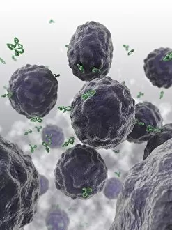
Antibodies and B cells. Computer artwork showing antibodies (green) neutralising cytokine activity in B cell (large, round) white blood cells

Background imageWhite Blood Cell Collection: Antibodies and B cells, artwork C016 / 6254

Antibodies and B cells, artwork C016 / 6254
Antibodies and B cells. Computer artwork showing antibodies (green) neutralising cytokine activity in B cell (large, round) white blood cells

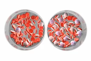
Background imageWhite Blood Cell Collection: Normal and leukaemic blood, artwork C016 / 6261

Normal and leukaemic blood, artwork C016 / 6261
Normal and leukaemic blood. Computer artwork comparing the appearance of normal blood (left) and that affected by leukaemia (right)

Background imageWhite Blood Cell Collection: Blood clot, SEM C015 / 9608

Blood clot, SEM C015 / 9608
Blood clot. Coloured scanning electron micrograph (SEM) of a blood clot from the inner wall of the left ventricle of a heart

Background imageWhite Blood Cell Collection: Blood cells, light micrograph C015 / 7130

Blood cells, light micrograph C015 / 7130
Blood cells. Light micrograph of red blood cells (erythrocytes, red) and white blood cells (leucocytes, nuclei stained pink)

Background imageWhite Blood Cell Collection: Neutrophil white blood cell, illustration C018 / 0741

Neutrophil white blood cell, illustration C018 / 0741
Neutrophil white blood cell, illustration. Neutrophils are the most abundant white blood cell in humans. They have a multi-lobed nucleus (dark purple)

Background imageWhite Blood Cell Collection: Budding HIV particles, TEM C018 / 0125

Budding HIV particles, TEM C018 / 0125
Budding HIV particles. Coloured transmision electron micrograph (TEM) of human immunodeficiency virus (HIV) particles (green) budding from the surface of a T lymphocyte (orange)

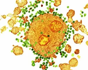
Background imageWhite Blood Cell Collection: Budding HIV particles, TEM C018 / 0124

Budding HIV particles, TEM C018 / 0124
Budding HIV particles. Coloured transmision electron micrograph (TEM) of human immunodeficiency virus (HIV) particles (gren) budding from the surface of a T lymphocyte (orange) from the H9 cell line

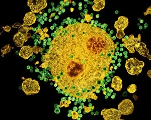
Background imageWhite Blood Cell Collection: Budding HIV particles, TEM C018 / 0123

Budding HIV particles, TEM C018 / 0123
Budding HIV particles. Coloured transmision electron micrograph (TEM) of human immunodeficiency virus (HIV) particles (blue) budding from the surface of a T lymphocyte (red) from the H9 cell line

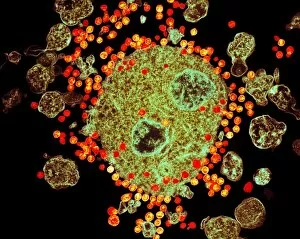
Background imageWhite Blood Cell Collection: Budding HIV particles, TEM C018 / 0127

Budding HIV particles, TEM C018 / 0127
Budding HIV particles. Coloured transmision electron micrograph (TEM) of human immunodeficiency virus (HIV) particles (orange) budding from the surface of a T lymphocyte (brown)

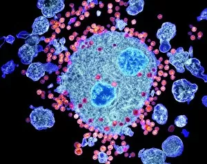
Background imageWhite Blood Cell Collection: Budding HIV particles, TEM C018 / 0126

Budding HIV particles, TEM C018 / 0126
Budding HIV particles. Coloured transmision electron micrograph (TEM) of human immunodeficiency virus (HIV) particles (pink) budding from the surface of a T lymphocyte (blue) from the H9 cell line

Background imageWhite Blood Cell Collection: Blood cells, illustration C018 / 0802

Blood cells, illustration C018 / 0802
Blood cells. All cellular blood components originate from the same cell, the haematopoietic stem cell. The stem cell differentiates into two types of progenitor cells

Background imageWhite Blood Cell Collection: Human immune response molecule complex

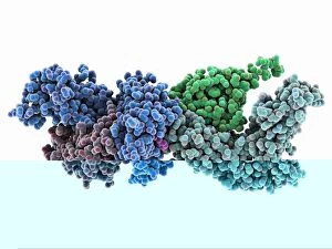
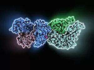
Human immune response molecule complex. Molecular model showing a human T-cell receptor and an HLA-A leukocyte (white blood cell) antigen bound to a TAX peptide from a virus

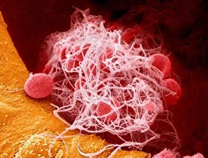
Background imageWhite Blood Cell Collection: Blood clot, SEM C016 / 9744

Blood clot, SEM C016 / 9744
Blood clot, coloured scanning electron micrograph (SEM). Red blood cells (erythrocytes) are trapped within a fibrin protein mesh (beige)

Background imageWhite Blood Cell Collection: Microglial white blood cell, SEM C019 / 0247

Microglial white blood cell, SEM C019 / 0247
Microglial white blood cell, SEM

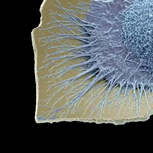
Background imageWhite Blood Cell Collection: Microglial white blood cell, SEM C019 / 0246

Microglial white blood cell, SEM C019 / 0246
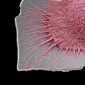
Microglia. Coloured scanning electron micrograph (SEM) of an activated microglial white blood cell. This immune system cell is found in the brain and spinal cord

Background imageWhite Blood Cell Collection: Microglial white blood cell, SEM C019 / 0245

Microglial white blood cell, SEM C019 / 0245
Microglia. Coloured scanning electron micrograph (SEM) of an activated microglial white blood cell. This immune system cell is found in the brain and spinal cord

Background imageWhite Blood Cell Collection: Human immune response molecule complex C014 / 0871

Human immune response molecule complex C014 / 0871
Human immune response molecule complex. Molecular model showing a human T-cell receptor and an HLA-A leukocyte (white blood cell) antigen bound to a TAX peptide from a virus

Background imageWhite Blood Cell Collection: Monocyte white blood cell, TEM

Monocyte white blood cell, TEM
Monocyte white blood cell. Transmission electron micrograph (TEM) of a section through a monocyte white blood cell, showing the cell nucleus (blue)

Background imageWhite Blood Cell Collection: HIV invading white blood cell, artwork

HIV invading white blood cell, artwork
HIV invading white blood cell. Computer artwork showing HIV (human immunodeficiency virus) particles (virions, purple) invading a white blood cell (large, centre) in the human bloodstream

Background imageWhite Blood Cell Collection: Blood cells, SEM C016 / 8026

Blood cells, SEM C016 / 8026
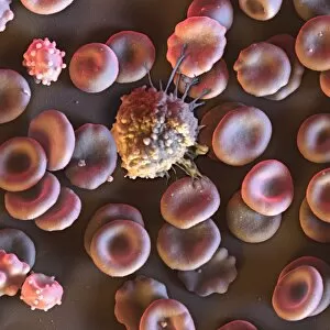
Blood cells. Coloured scanning electron micrograph (SEM) of a lymphocyte white blood cell (centre) and red blood cells (erythrocytes, circular)

Background imageWhite Blood Cell Collection: Activated macrophage, SEM C015 / 6377

Activated macrophage, SEM C015 / 6377
Activated macrophage. Coloured scanning electron micrograph (SEM) of an active macrophage white blood cell. Macrophages are cells of the bodys immune system that are found in the tissues rather than

Background imageWhite Blood Cell Collection: Activated macrophage, SEM C015 / 6376

Activated macrophage, SEM C015 / 6376
Activated macrophage. Coloured scanning electron micrograph (SEM) of an active macrophage white blood cell. Macrophages are cells of the bodys immune system that are found in the tissues rather than


All Professionally Made to Order for Quick Shipping

"Guardians of Health: Exploring the Mighty White Blood Cell" T lymphocytes and cancer cells: Unveiling the battle within, as T lymphocytes wage war against cancer cells, SEM C001 / 1679. Neutrophil engulfing MRSA: Witness the incredible defense mechanism as a neutrophil engulfs MRSA bacteria, SEM C018 / 8596. Dendritic cells artwork: Unlocking the secrets of immune response with stunning artwork depicting dendritic cells at work. TEM of human white blood cell bearing HLA antigen: Peering into the intricate world of immunity through a transmission electron microscope image showcasing a white blood cell displaying HLA antigens. Blood cells in harmony: A mesmerizing glimpse into our life force - an ensemble of diverse blood cells working together for our well-being. Coloured SEM of a white blood cell (lymphocyte): Dive deep into the vibrant realm of lymphocytes captured in this captivating colored scanning electron microscope image. Basophil white blood cell: Discovering the lesser-known heroes among us - basophilic white blood cells that play crucial roles in allergic reactions and inflammation. Bacteria infecting a macrophage, SEM: Witness how macrophages confront invading bacteria head-on under high-resolution scanning electron microscopy imagery. Blood coagulation cascade artwork: Unraveling the intricacies behind clot formation with an artistic representation capturing every step in this vital process, artwork C016 / 9873 Red and white blood cells, SEM : Marvel at nature's palette as red and white blood cells come alive under scanning electron microscopy - an exquisite symphony within us all. Blood clot, SEM C016 / 9747 : Delving into hemostasis marvels with a striking scanning electron micrograph revealing intricate details within a formed clot structure. Dohle bodies in blood cell, micrograph.