
White Blood Cells Collection
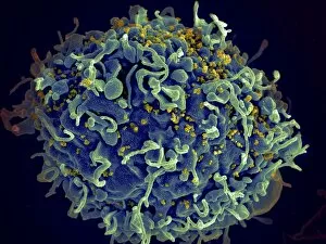
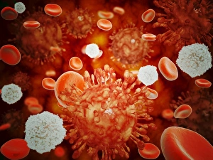
"Exploring the Mighty Defenders: Unveiling the World of White Blood Cells" Witness the battle against HIV as captured in a stunning scanning electron micrograph


All Professionally Made to Order for Quick Shipping
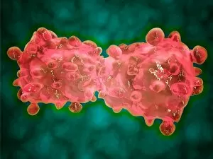
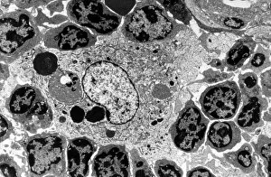
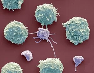
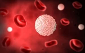
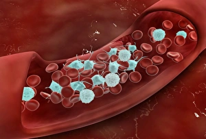
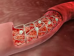
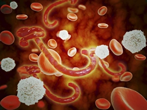
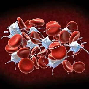
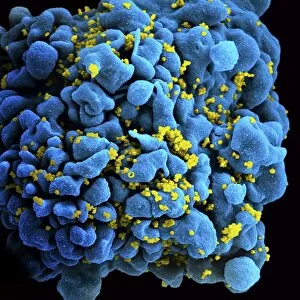
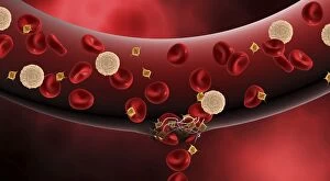
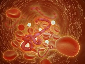
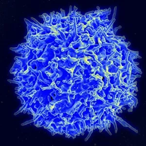
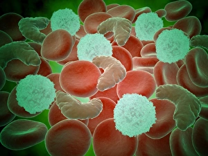
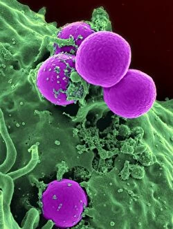
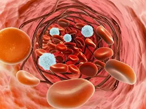
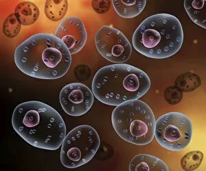
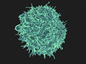
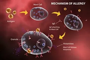
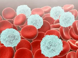
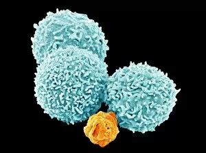
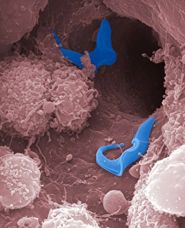
"Exploring the Mighty Defenders: Unveiling the World of White Blood Cells" Witness the battle against HIV as captured in a stunning scanning electron micrograph, revealing white blood cells valiantly fighting off infection within a human H9 T cell. A captivating conceptual image showcases the intricate collaboration between platelets, red blood cells, and white blood cells - our body's ultimate defense squad. Delve into the microscopic world to observe an intense view of leukemia cells, reminding us of the crucial role white blood cells play in combating cancerous invaders. Discover the remarkable teamwork between macrophages and lymphocytes through a striking transmission electron microscope image that unveils their strategic alliance against pathogens. Immerse yourself in an extraordinary SEM capture displaying white blood cells and platelets working together harmoniously to maintain our body's delicate balance - SEM C016 / 3099. Marvel at another mesmerizing SEM image (SEM C016 / 3098) showcasing how white blood cells and platelets collaborate seamlessly to safeguard our health from potential threats. Get up close with lymphocytes inside a hair follicle through a fascinating scanning electron microscope view, highlighting their presence throughout various parts of our body's defense system. Peer into the microscopic realm once again to witness human B-cells in action – these specialized they can key players in adaptive immunity, ensuring long-term protection against infections. Explore an astonishing microscopic view capturing bustling activity as white blood cells navigate within a blood vessel – constantly patrolling for any signs of danger lurking within our circulatory system. Observe with awe as clotting occurs inside an artery through this captivating microscopic perspective; it serves as a reminder that without efficient functioning white blood cells, such events could pose severe risks to our health.