Fraud Blocker Skip to main content

Biomedical Collection

"Exploring the Biomedical Frontier

Background imageBiomedical Collection: United Kingdom, England, London, Kings Cross. The Francis Crick Institute

United Kingdom, England, London, Kings Cross. The Francis Crick Institute - biomedical research centre in London, with St. Pancras railway station in the rear of shot

Background imageBiomedical Collection: Biomedical centre, London -- a woman attaches sensors to a patient. Date: 1972

Biomedical centre, London -- a woman attaches sensors to a patient. Date: 1972

Background imageBiomedical Collection: Biomedical centre, London -- a woman in an office operates a telex machine

Biomedical centre, London -- a woman in an office operates a telex machine. Date: 1972

Background imageBiomedical Collection: Biomedical centre, London -- a patient in a locked room listens to something through

Biomedical centre, London -- a patient in a locked room listens to something through headphones while a woman operates a machine in the room beyond. Date: 1972

Background imageBiomedical Collection: Biomedical centre, London -- a woman operates a machine while a patient in a locked room

Biomedical centre, London -- a woman operates a machine while a patient in a locked room listens to something through headphones. Date: 1972

Background imageBiomedical Collection: Nucleic acid, illustration

Nucleic acid, illustration

Background imageBiomedical Collection: A female nude from the waist up with a doctors hands conducting a clinical breast

A female nude from the waist up with a doctors hands conducting a clinical breast examination

Background imageBiomedical Collection: Illustration showing upper and lower limbs

Illustration showing upper and lower limbs
Medicine: bones. Upper and lower limbs. Drawing

Background imageBiomedical Collection: Scanning electron micrograph of HIV particles infecting a human H9 T cell

Scanning electron micrograph of HIV particles infecting a human H9 T cell

Background imageBiomedical Collection: Coxiella burnetii, the bacteria that causes Q Fever

Coxiella burnetii, the bacteria that causes Q Fever. A dry fracture of a Vero cell exposing the contents of a vacuole where Coxiella burnetii are busy growing

Background imageBiomedical Collection: Kaposis sarcoma on the skin of an AIDS patient

Kaposis sarcoma on the skin of an AIDS patient

Background imageBiomedical Collection: Lasers used to position a patient wearing a short face mask in a computed tomography

Lasers used to position a patient wearing a short face mask in a computed tomography
Lasers are used to position a patient model wearing a short face mask in a computed tomography (CT) scanner to obtain images of a tumor before radiation therapy

Background imageBiomedical Collection: Brazil - Sao Paulo - Serpentarium of the Instituto Butantan

Brazil - Sao Paulo - Serpentarium of the Instituto Butantan, a Brazilian biomedical research centre affiliated to the Sao Paulo State Secretary of Health

Background imageBiomedical Collection: Illustration showing muscular system of human head

Illustration showing muscular system of human head
Medicine: Human anatomy, muscular system, head. Muscles, nerves, glands. Drawing

Background imageBiomedical Collection: Biomedical centre, London

Biomedical centre, London -- a woman attaches sensors to a patient. Date: 1972

Background imageBiomedical Collection: Researcher uses pipette in lab

Researcher uses pipette in lab

Background imageBiomedical Collection: Red blood cells on an agar plate are used to diagnose infection

Red blood cells on an agar plate are used to diagnose infection. The plate on the left shows a positive staphyloccus infection

Background imageBiomedical Collection: Rack of test tubes

Rack of test tubes

Background imageBiomedical Collection: Large eroded plaque on the leg with areas of crust

Large eroded plaque on the leg with areas of crust
Metastatic Melanoma. Large eroded plaque on the leg with areas of crust. Scattered eroded dermal nodules are also present

Background imageBiomedical Collection: Melanoma on a patients skin

Melanoma on a patients skin

Background imageBiomedical Collection: Colorized image of HIV-infected H9 T-cell

Colorized image of HIV-infected H9 T-cell

Background imageBiomedical Collection: Bacteria from human skin grown on agar in the laboratory

Bacteria from human skin grown on agar in the laboratory

Background imageBiomedical Collection: 3D structure of melanoma cell

3D structure of melanoma cell
3D structure of a melanoma cell derived by ion abrasion scanning electron microscopy

Background imageBiomedical Collection: Automated DNA sequencing laboratory equipment and paraphernalia

Automated DNA sequencing laboratory equipment and paraphernalia

Background imageBiomedical Collection: Scanning electron micrograph of just-divided HeLa cells

Scanning electron micrograph of just-divided HeLa cells. Zeiss Merlin HR-SEM

Background imageBiomedical Collection: Multiphoton fluorescence image of HeLa cells

Multiphoton fluorescence image of HeLa cells with cytoskeletal microtubules (magenta) and DNA (cyan)

Background imageBiomedical Collection: A colorized scanning electron micrograph of a white blood cell eating MRSA

A colorized scanning electron micrograph of a white blood cell eating MRSA
A colorized scanning electron micrograph of a white blood cell eating an antibiotic resistant strain of Staphylococcus aureus bacteria, commonly known as MRSA

Background imageBiomedical Collection: Scanning electron micrograph of a human T cell

Scanning electron micrograph of a human T cell
Scanning electron micrograph of a human T lymphocyte (also called a T cell) from the immune system of a healthy donor

Background imageBiomedical Collection: Vials containing chemicals in research lab

Vials containing chemicals in research lab

Background imageBiomedical Collection: Radiation therapists fit a short face mask to a patient during radiation therapy

Radiation therapists fit a short face mask to a patient during radiation therapy
Two radiation therapists fit a short face mask to a patient model to steady the head during radiation therapy

Background imageBiomedical Collection: Scanning electron micrograph of a human neutrophil ingesting MRSA

Scanning electron micrograph of a human neutrophil ingesting MRSA

Background imageBiomedical Collection: An automatic spinning device to mix tissue culture medium

An automatic spinning device to mix tissue culture medium. Tissue culture medium provides nourishment to growing cells

Background imageBiomedical Collection: 3D structure of pyruvate dehydrogenase complex

3D structure of pyruvate dehydrogenase complex
3D structure of the 11 megadalton pyruvate dehydrogenase complex which is responsible for synthesis of the key metabolite acetyl CoA

Background imageBiomedical Collection: Scanning electron micrograph of Staphylococcus and a dead human neutrophil

Scanning electron micrograph of Staphylococcus and a dead human neutrophil
Scanning electron micrograph of methicillin-resistant Staphylococcus aureus and a dead human neutrophil

Background imageBiomedical Collection: Generic vials with chemicals in research lab

Generic vials with chemicals in research lab

Background imageBiomedical Collection: Colorized scanning electron micrograph of a T lymphocyte

Colorized scanning electron micrograph of a T lymphocyte

Background imageBiomedical Collection: Primary cultures of superior cervical ganglia cells

Primary cultures of superior cervical ganglia cells

Background imageBiomedical Collection: Researcher in lab uses pipette and measures RNA

Researcher in lab uses pipette and measures RNA

Background imageBiomedical Collection: Field of blood cells illustration

Field of blood cells illustration
This is a field of blood cells. The bi-concave disks are red blood cells or erythrocytes. The white cell with the dark purplish, multi-lobed nucleus is a neutrophil

Background imageBiomedical Collection: Installing a flow cell on an Illumina cluster station

Installing a flow cell on an Illumina cluster station

Background imageBiomedical Collection: Staphylococcus Bacteria from human skin grown on agar

Staphylococcus Bacteria from human skin grown on agar
Staphylococcus bacteria from human skin grown on agar in the laboratory

Background imageBiomedical Collection: Patient positioned in computed tomography scanner

Patient positioned in computed tomography scanner
A patient model wearing a short face mask is positioned in a computed tomography (CT) scanner to obtain images of a tumor before radiation therapy

Background imageBiomedical Collection: Scanning electron micrograph of a schistosome parasite

Scanning electron micrograph of a schistosome parasite, which enters the body through the skin of persons coming in contact with infested waters. The adult worm lives in the veins of its host

Background imageBiomedical Collection: Structure of HIV

Structure of HIV

Background imageBiomedical Collection: Neurofibromatosis, a genetic disorder of the nervous system

Neurofibromatosis, a genetic disorder of the nervous system
A genetic disorder of the nervous system, neurofibromatosis causes tumors to form on nerves throughout the body, including a type of tumor called an optic nerve glioma that can result in childhood

Background imageBiomedical Collection: Colorized scanning electron micrograph of filamentous Ebola virus particles

Colorized scanning electron micrograph of filamentous Ebola virus particles (blue) budding from a chronically infected VERO E6 cell (yellow-green)

Background imageBiomedical Collection: Rack of pipettes

Rack of pipettes

Background imageBiomedical Collection: Artistic rendering of the surface of a human dendritic cell

Artistic rendering of the surface of a human dendritic cell illustrating the unexpected discovery of sheet-like processes that fold back onto the membrane surface


All Professionally Made to Order for Quick Shipping

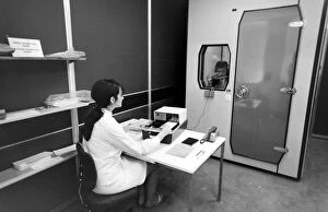
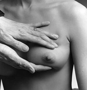
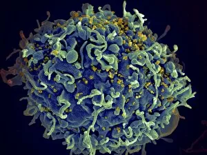
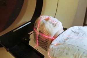
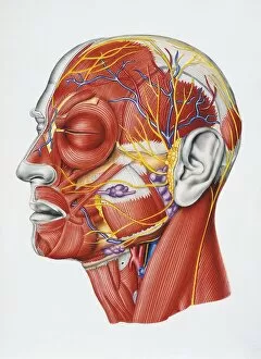
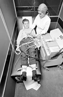
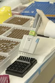
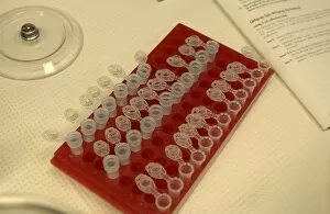
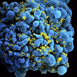
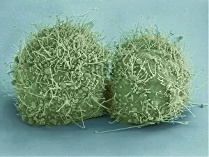
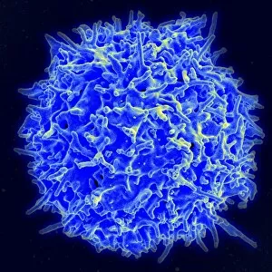
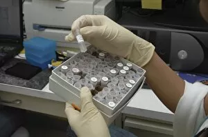
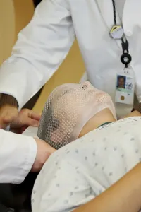
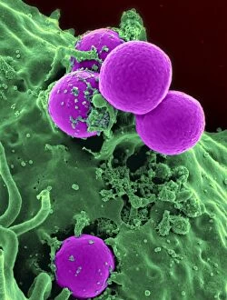
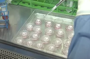
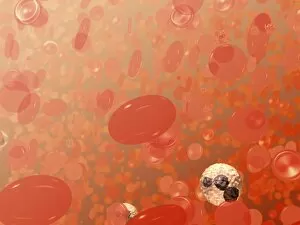
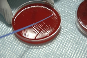
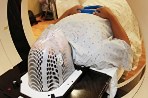
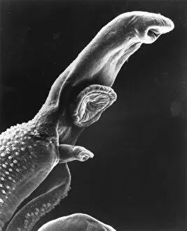
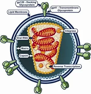
"Exploring the Biomedical Frontier: Unveiling London's Pioneering Work at Kings Cross" Step into the captivating world research as we journey through the heart of United Kingdom's scientific hub, London. Nestled in the vibrant district of Kings Cross lies The Francis Crick Institute, a beacon for groundbreaking discoveries and advancements in healthcare. Transporting us back to 1972, an era marked by remarkable progress, we witness a dedicated woman attaching sensors to a patient at the Biomedical Centre. With precision and care, she sets the stage for cutting-edge experiments that would shape medical history. In another corner of this bustling center, amidst rows of buzzing machines and diligent researchers, a woman operates a telex machine with unwavering focus. This intricate communication system serves as a vital lifeline connecting scientists across borders—a testament to collaboration transcending boundaries. Venturing further within these walls reveals locked rooms where patients eagerly listen to something transformative—perhaps an innovative treatment or life-changing breakthroughs whispered through hidden speakers. Their hope intertwines with relentless efforts behind closed doors; their stories interwoven with countless hours spent unraveling mysteries within these hallowed halls. As our exploration continues, we encounter yet another determined woman operating an advanced machine designed to test patients' hearing capabilities. Through meticulous analysis and compassionate care, she paves new avenues for understanding auditory health—an invaluable contribution towards enhancing lives. Amidst it all stands Nucleic Acid—an illustration symbolizing genetic blueprints that hold immense potential for unlocking cures and deciphering human existence itself. It embodies the essence research—the pursuit of knowledge intertwined with compassion-driven innovation. The Biomedical Centre becomes our gateway into this extraordinary realm where science meets humanity—a place where tireless dedication converges with boundless curiosity. Each room holds untold stories waiting to be discovered; each researcher is driven by an unyielding passion to make a difference in people's lives.