
Knee Joint Collection
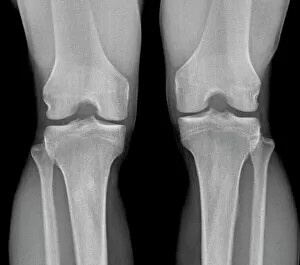
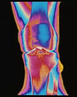
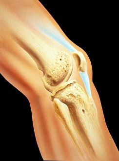
"The intricate beauty of a normal knee joint revealed through an X-ray, showcasing its remarkable structure and functionality


All Professionally Made to Order for Quick Shipping
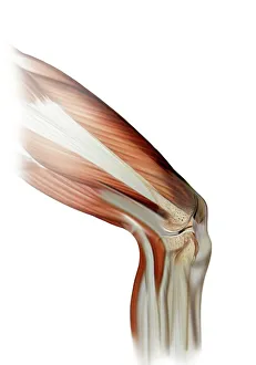
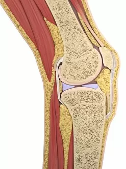
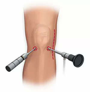
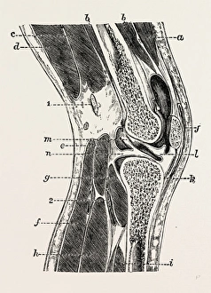
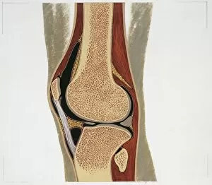
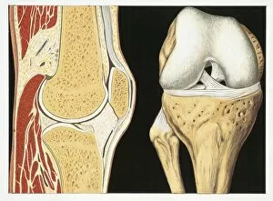
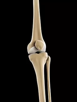
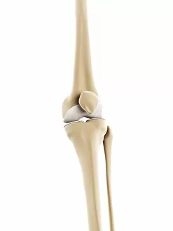
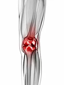
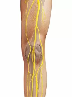
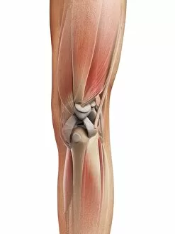
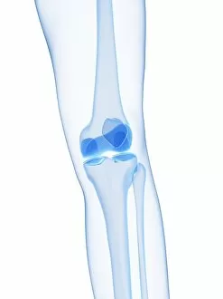
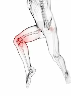
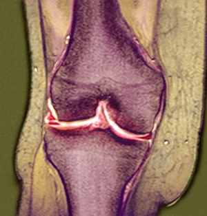
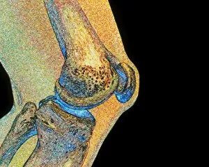
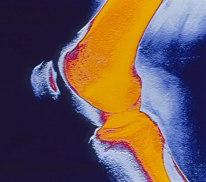
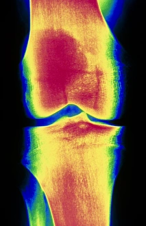
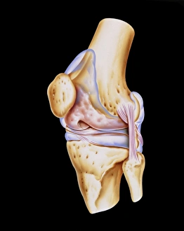
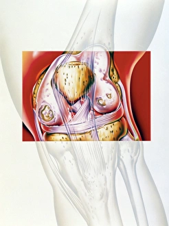
"The intricate beauty of a normal knee joint revealed through an X-ray, showcasing its remarkable structure and functionality. " "Comparing a healthy knee joint to an X-ray image, highlighting the importance of maintaining optimal joint health. " "A damaged knee ligament depicted through captivating artwork, emphasizing the vulnerability of this crucial joint. " "Exploring the vibrant hues of a colored X-ray capturing the complexity and vitality of a human knee joint. " "The perils faced by runners showcased in thought-provoking conceptual artwork, shedding light on common running injuries that affect the knee. " "Glimpsing into the inner workings of a normal lower leg with an X-ray, focusing on the pivotal role played by a well-functioning knee joint. " "Dive into detailed artwork depicting the intricate anatomy of a knee joint, unraveling its fascinating composition and mechanics. " "Unveiling arthroscopic surgical repair as an innovative solution for restoring functionality to injured knees, offering hope for those facing mobility challenges. " "A glimpse at perfection - behold a flawless depiction of a normal knee captured in an awe-inspiring X-ray image. "Delving into history with Abraham de Visscher's 1605-67 Amsterdam merchant who contributed to our understanding of vertical sections within the complex realm of knee-joint exploration. " "Illuminating medical equipment and surgical instruments used throughout history to study and treat various conditions affecting the delicate structures within our knees. " "Intricate illustration revealing every detail and nuance present within one's own unique kneecap; marvel at nature's design.