Jigsaw Puzzle : Ant foot, SEM


![]()
Jigsaw Puzzles from Science Photo Library
Ant foot, SEM
Ant foot. Coloured scanning electron micrograph (SEM) of the tip of a leg from an ant (family Formicidae). The end of an insect leg consists of the final segment, which is called the tarsus. The tarsus itself has several segments, and here ends in a pretarsus that has two claws (orange). Leg bristles and hair-like sensory structures are also seen
Science Photo Library features Science and Medical images including photos and illustrations
Media ID 6461404
© SUSUMU NISHINAGA/SCIENCE PHOTO LIBRARY
Bristle Bristles Claw Claws False Colour Foot Hair Hairs Segment Sense Sensory Tarsus False Coloured
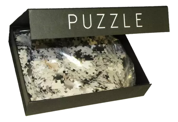
Jigsaw Puzzle (500 Pieces)
Discover the intricacies of the natural world with Media Storehouse's Jigsaw Puzzles. Our latest addition to the collection is an awe-inspiring puzzle featuring an Ant Foot SEM image from Science Photo Library. This high-definition puzzle showcases the stunning details of an ant's leg tip in vibrant colors, captured through Scanning Electron Microscopy. Challenge yourself and your family to piece together this captivating jigsaw puzzle, perfect for hours of educational and entertaining fun. Delve deeper into the intricacies of the ant's anatomy and marvel at the beauty of the microscopic world.
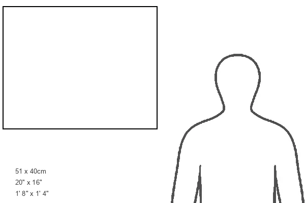
500 piece puzzles are custom made in Australia and hand-finished on 100% recycled 1.6mm thick laminated puzzle boards. There is a level of repetition in jigsaw shapes with each matching piece away from its pair. The completed puzzle measures 40x51cm and is delivered packaged in an attractive presentation box specially designed to fit most mail slots with a unique magnetic lid
Jigsaw Puzzles are an ideal gift for any occasion
Estimated Product Size is 50.7cm x 40.3cm (20" x 15.9")
These are individually made so all sizes are approximate
Artwork printed orientated as per the preview above, with landscape (horizontal) or portrait (vertical) orientation to match the source image.
EDITORS COMMENTS
This print showcases the intricate details of an ant's foot, captured using a scanning electron microscope (SEM). The image reveals the remarkable complexity of an insect leg, specifically focusing on the tarsus - the final segment. Within the tarsus, multiple segments can be observed, leading to a pretarsus adorned with two vibrant orange claws. The photograph also highlights various sensory structures and bristles that cover the leg. These hair-like features play a crucial role in helping ants navigate their surroundings and detect changes in their environment. Nature enthusiasts and zoology aficionados will find this print fascinating as it provides a close-up glimpse into the world of these tiny creatures. It serves as a reminder of how diverse and extraordinary life forms can be even at microscopic levels. With its false-colored presentation, this SEM image beautifully combines artistry with scientific precision. The vivid hues add depth to each element while emphasizing their significance within ant anatomy. Science Photo Library has once again delivered an awe-inspiring visual representation that bridges science and aesthetics seamlessly. This print is not only visually striking but also serves as a testament to our ever-growing understanding of nature's intricacies through advanced imaging techniques like scanning electron microscopy.
MADE IN AUSTRALIA
Safe Shipping with 30 Day Money Back Guarantee
FREE PERSONALISATION*
We are proud to offer a range of customisation features including Personalised Captions, Color Filters and Picture Zoom Tools
SECURE PAYMENTS
We happily accept a wide range of payment options so you can pay for the things you need in the way that is most convenient for you
* Options may vary by product and licensing agreement. Zoomed Pictures can be adjusted in the Cart.