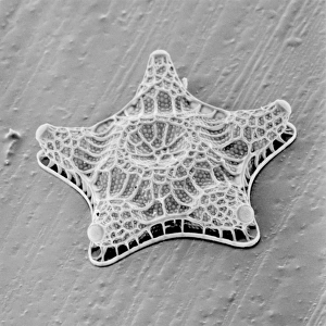
Actinoptychus, diatom

![]()
Wall Art and Photo Gifts from Mary Evans Picture Library
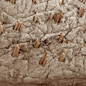
Actinoptychus, diatom
Scanning electron microscope image of the exterior valve of the diatom Actinoptychus (x 500 on a standard 9 cm wide print)
Mary Evans Picture Library makes available wonderful images created for people to enjoy over the centuries
Media ID 8580582
© Mary Evans Picture Library 2015 - https://copyrighthub.org/s0/hub1/creation/maryevans/MaryEvansPictureID/10707859
Alga Algae Algal Bacillariophyceae Chromalveolata Chromista Diatom Electron Micrograph Eukaryote Eukaryotic Micrograph Microscope Image Ochrophyta Protist Protista Scanning Electron Micrograph Scanning Electron Microscope Scanning Electron Microscope Image Sem Image Shell Silica Valve
EDITORS COMMENTS
1. Title: Ornate Diatom Actinoptychus: A Magnificent Display of Silica Architecture Revealed through Scanning Electron Microscopy 2.. This stunning scanning electron microscope image (SEM) showcases the intricately ornamented exterior valve of the diatom Actinoptychus, a microscopic alga belonging to the class Bacillariophyceae within the phylum Chromista and the kingdom Protista. Diatoms are unicellular eukaryotes, renowned for their unique and beautiful silica shells, which provide structural support and protection. Actinoptychus is a member of the Coscinodiscophyceae, specifically the Coscinodiscales and the family Heliopeltidae. The valve in this image displays an impressive array of radial and circular ornamentations, which are characteristic of this diatom species. The intricate patterns are formed by the deposition of silica, a process that occurs through the interaction of the diatom with specific minerals in its environment. The SEM image provides a captivating glimpse into the microscopic world of Actinoptychus and its silica shell. This ornate diatom is a testament to the intricacy and diversity of life at the microscopic level, highlighting the beauty and complexity found in the smallest of organisms. This image was captured using a scanning electron microscope at a magnification of 500x, providing a detailed view of the diatom's intricate valve structure. The image is an essential resource for researchers studying diatoms and their role in aquatic ecosystems, as well as for those interested in the wonders of the microscopic world. In conclusion, the Actinoptychus diatom, as depicted in this SEM image, is a remarkable example of the beauty and complexity found in the microscopic world. Its intricately ornamented silica shell serves as a testament to the intricacy and diversity of life at the smallest scales, offering a fascinating glimpse into the unseen world of diatoms and their role in aquatic ecosystems.
MADE IN AUSTRALIA
Safe Shipping with 30 Day Money Back Guarantee
FREE PERSONALISATION*
We are proud to offer a range of customisation features including Personalised Captions, Color Filters and Picture Zoom Tools
FREE COLORIZATION SERVICE
You can choose advanced AI Colorization for this picture at no extra charge!
SECURE PAYMENTS
We happily accept a wide range of payment options so you can pay for the things you need in the way that is most convenient for you
* Options may vary by product and licensing agreement. Zoomed Pictures can be adjusted in the Cart.