Mouse Mat : Coloured SEM of human striated muscle

![]()
Home Decor from Science Photo Library
Coloured SEM of human striated muscle
Striated muscle. Coloured scanning electron micrograph (SEM) of human striated muscle. Striated muscle occurs in all skeletal muscles and its movement is under voluntary control. It is composed of long muscle fibres made up of cylindrical myofibrils (green) containing contractile proteins. The parallel arrangement of the myo- fibrils is seen here. The regular pattern of the contractile proteins in the myofibrils produces characteristic stripes or striations (not seen). Infiltrating the myofibrils at these regular intervals is sarcoplasmic reticulum (globose, orange), the muscles specialized membrane system. Magnification: unknown
Science Photo Library features Science and Medical images including photos and illustrations
Media ID 6420310
© STEVE GSCHMEISSNER/SCIENCE PHOTO LIBRARY
Muscles Myofibril Myofibrils Sarcoplasmic Reticulum Skeletal Skeletal Muscle Striated Muscle
Mouse Pad
Bring some life into your office, or create a heartfelt gift, with a personalised deluxe Mouse Mat. Made of high-density black foam with a tough, stain-resistant inter-woven cloth cover they will brighten up any home or corporate office.
Archive quality photographic print in a durable wipe clean mouse mat with non slip backing. Works with all computer mice
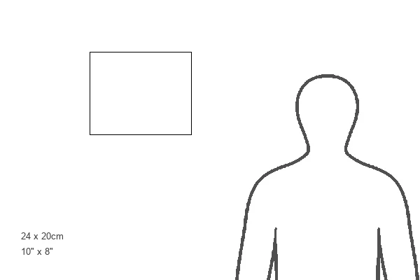
Estimated Product Size is 24.2cm x 19.7cm (9.5" x 7.8")
These are individually made so all sizes are approximate
Artwork printed orientated as per the preview above, with landscape (horizontal) or portrait (vertical) orientation to match the source image.
EDITORS COMMENTS
This print showcases a coloured scanning electron micrograph (SEM) of human striated muscle, providing an intricate glimpse into the fascinating world of anatomy. Striated muscle is found in all skeletal muscles and is responsible for voluntary movement. The image reveals long muscle fibres composed of cylindrical myofibrils, which are highlighted in a vibrant green hue. These myofibrils contain contractile proteins that enable muscular contraction. The parallel arrangement of the myofibrils creates a visually striking pattern, reminiscent of delicate stripes or striations. However, these distinct markings are not visible in this particular SEM capture. Intriguingly, infiltrating the myofibrils at regular intervals is the sarcoplasmic reticulum depicted as globose structures glowing orange. This specialized membrane system plays a crucial role in supporting muscle function. While the exact magnification remains unknown, this photograph provides an extraordinary level of detail that allows us to appreciate the complexity and beauty inherent within our own bodies. It serves as a reminder of the incredible intricacies that exist within our skeletal muscles and how they work harmoniously to facilitate movement. This remarkable image from Science Photo Library offers both scientific insight and aesthetic appeal, making it an ideal addition to any collection exploring topics such as muscle anatomy, skeletal structure, or human physiology without mentioning commercial use or specific companies involved with its production.
MADE IN AUSTRALIA
Safe Shipping with 30 Day Money Back Guarantee
FREE PERSONALISATION*
We are proud to offer a range of customisation features including Personalised Captions, Color Filters and Picture Zoom Tools
SECURE PAYMENTS
We happily accept a wide range of payment options so you can pay for the things you need in the way that is most convenient for you
* Options may vary by product and licensing agreement. Zoomed Pictures can be adjusted in the Cart.