Photo Mug > Fine Art Storehouse > Science Inspired Art
Photo Mug : Mushroom gill, LM


![]()
Home Decor from Fine Art Storehouse
Mushroom gill, LM
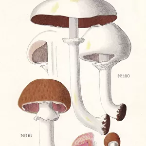
Mushroom gills. High power light micrograph (LM) of a section through the gills of a mushroom, Agaricus sp. (formerly Psalliota sp.). The hymenial layer, or hymenium, produces the spore-bearing structures known as basidia (red). Many spores (basidiospores) are visible at the surface of this layer (yellow-black)
Unleash your creativity and transform your space into a visual masterpiece!
STEVE GSCHMEISSNER/SCIENCE PHOTO
Media ID 19527383
© Science Photo Library
Biologica Biology Horizontal Image Scientific
Photo Mug
Brighten up your mornings with our Media Storehouse Photo Mugs, featuring stunning high power light micrograph images from the Fine Art Storehouse. This particular design showcases the intricate detail of Mushroom gills, as captured by Steve Gschmeissner in his scientific exploration of the Agaricus sp. (formerly Psalliota sp.) mushroom. Each mug holds your favorite beverage and serves as a daily reminder of the beauty and complexity of the natural world. Perfect for the nature lover or scientist in your life.
A personalised photo mug blends sentimentality with functionality, making an ideal gift for cherished loved ones, close friends, or valued colleagues. Preview may show both sides of the same mug.
Elevate your coffee or tea experience with our premium white ceramic mug. Its wide, comfortable handle makes drinking easy, and you can rely on it to be both microwave and dishwasher safe. Sold in single units, preview may show both sides of the same mug so you can see how the picture wraps around.
Mug Size is 8.1cm high x 9.6cm diameter (3.2" x 3.8")
These are individually made so all sizes are approximate
FEATURES IN THESE COLLECTIONS
> Fine Art Storehouse
> Science Inspired Art
> SEM (Scanning Electron Microscope)
> Fine Art Storehouse
> Science Inspired Art
EDITORS COMMENTS
This print showcases the intricate beauty of a mushroom gill, captured under high power light micrograph (LM). The image reveals a section through the gills of an Agaricus sp. mushroom, formerly known as Psalliota sp. The mesmerizing hymenial layer, or hymenium, takes center stage in this composition. It is responsible for producing the striking spore-bearing structures called basidia, which are depicted in vibrant red hues. Upon closer examination, numerous basidiospores can be observed at the surface of this remarkable layer. These tiny yellow-black specks add depth and texture to the overall visual narrative presented by this photograph. The horizontal orientation of the image allows viewers to fully immerse themselves in its scientific wonder. Its inclusion within biology and scientific contexts makes it an ideal addition to educational materials or research publications. Renowned photographer Steve Gschmeissner skillfully captures nature's intricacies with his lens, transforming them into works of art that bridge science and aesthetics seamlessly. This particular piece from Fine Art Storehouse exemplifies Gschmeissner's ability to showcase both technical precision and artistic vision simultaneously. Whether displayed on a gallery wall or used as an educational tool, this stunning photograph invites viewers into the fascinating world of mushrooms while highlighting their biological significance.
MADE IN AUSTRALIA
Safe Shipping with 30 Day Money Back Guarantee
FREE PERSONALISATION*
We are proud to offer a range of customisation features including Personalised Captions, Color Filters and Picture Zoom Tools
SECURE PAYMENTS
We happily accept a wide range of payment options so you can pay for the things you need in the way that is most convenient for you
* Options may vary by product and licensing agreement. Zoomed Pictures can be adjusted in the Cart.