Photo Mug > Science > SEM
Photo Mug : Red blood cells and platelets, SEM


![]()
Home Decor from Fine Art Storehouse
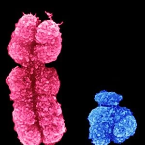
Red blood cells and platelets, SEM
Red blood cells and platelets. Coloured scanning electron micrograph (SEM) of human erythrocytes (red blood cells) and a platelet aggregate (orange). Platelets are fragments of white blood cells that under normal circumstances are small and biconcave in form. However, if there is a break in the surface of a blood vessel the platelets come into contact with molecules they are not used to and become activated. They become amorphous in form, with long projections (pseudopodia) that help them adhere to other cells and each other, forming a clot. Magnification: x3000 when printed at 10 centimetres wide
Unleash your creativity and transform your space into a visual masterpiece!
STEVE GSCHMEISSNER/SCIENCE PHOTO
Media ID 18106607
© Science Photo Library
3 Dimensional Anatomical Anatomy Biological Biology Biconcave
Photo Mug
Bring the wonders of the microscopic world into your daily life with our Media Storehouse Photo Mugs. Featuring an awe-inspiring image of red blood cells and platelets in vibrant detail, captured through the lens of STEVE GSCHMEISSNER's Scanning Electron Microscope. Each mug holds your favorite beverage while serving as a captivating conversation starter. Embrace the beauty of science in every sip.
A personalised photo mug blends sentimentality with functionality, making an ideal gift for cherished loved ones, close friends, or valued colleagues. Preview may show both sides of the same mug.
Elevate your coffee or tea experience with our premium white ceramic mug. Its wide, comfortable handle makes drinking easy, and you can rely on it to be both microwave and dishwasher safe. Sold in single units, preview may show both sides of the same mug so you can see how the picture wraps around.
Mug Size is 9.6cm high x 8.1cm diameter (3.8" x 3.2")
These are individually made so all sizes are approximate
FEATURES IN THESE COLLECTIONS
> Fine Art Storehouse
> Science Inspired Art
> SEM (Scanning Electron Microscope)
> Fine Art Storehouse
> Science Inspired Art
EDITORS COMMENTS
This print showcases the intricate world of our circulatory system. In this coloured scanning electron micrograph (SEM), we are granted a mesmerizing glimpse into the realm of red blood cells and platelets. The image reveals a cluster of human erythrocytes, or red blood cells, alongside an aggregate of platelets in a striking orange hue. Under normal circumstances, platelets exist as small and biconcave fragments derived from white blood cells. However, when there is damage to a blood vessel's surface, these remarkable cellular entities undergo activation. As they come into contact with unfamiliar molecules, their shape transforms dramatically. They assume an amorphous form with long projections known as pseudopodia that aid in adhering to other cells and each other. The result is the formation of a clot – nature's ingenious mechanism for preventing excessive bleeding and promoting wound healing. This SEM image captures the beauty within this vital process at an astonishing magnification level of x3000 when printed at 10 centimetres wide. Through this artwork by Steve Gschmeissner from Science Photo Library, we are reminded once again of the complexity and elegance inherent in our biological makeup. It serves as both a testament to scientific discovery and an artistic masterpiece that invites us to marvel at the wonders hidden within ourselves.
MADE IN AUSTRALIA
Safe Shipping with 30 Day Money Back Guarantee
FREE PERSONALISATION*
We are proud to offer a range of customisation features including Personalised Captions, Color Filters and Picture Zoom Tools
SECURE PAYMENTS
We happily accept a wide range of payment options so you can pay for the things you need in the way that is most convenient for you
* Options may vary by product and licensing agreement. Zoomed Pictures can be adjusted in the Cart.