Photo Mug : Choroid layer of the eye, SEM


![]()
Home Decor from Science Photo Library
Choroid layer of the eye, SEM
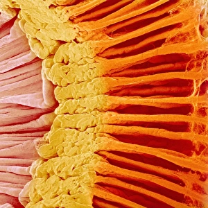
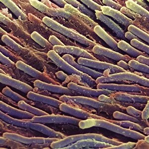
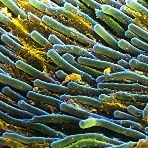
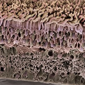
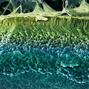
Choroid layer of the eye. Coloured scanning electron micrograph (SEM) of a section through the choroid layer of the eye. A pigment cell (dark brown) is at upper centre. The choroid layer lies behind the sclera (white of the eye) and in front of the retina. It is highly vascular and supplies blood to the back of the eye. It is pigmented to absorb excessive light, preventing internal reflections that would form multiple images on the retina. Magnification: x1100 when printed 10 centimetres wide
Science Photo Library features Science and Medical images including photos and illustrations
Media ID 6448703
© STEVE GSCHMEISSNER/SCIENCE PHOTO LIBRARY
Choroid False Colour Histological Histology Layer Pigmented Sense Sight Vascular Vision False Coloured
Photo Mug
Brighten up your morning routine with our Media Storehouse Photo Mugs, featuring the stunning scientific detail of the Choroid layer of the Eye. This captivating coloured Scanning Electron Micrograph (SEM) image from Science Photo Library showcases the intricate structure of this vital part of the eye, with a pigment cell proudly displayed in the upper centre. Each mug holds your favourite beverage, providing a daily reminder of the wonders of science as you take a sip. Make every drink a moment of discovery with Media Storehouse Photo Mugs.
A personalised photo mug blends sentimentality with functionality, making an ideal gift for cherished loved ones, close friends, or valued colleagues. Preview may show both sides of the same mug.
Elevate your coffee or tea experience with our premium white ceramic mug. Its wide, comfortable handle makes drinking easy, and you can rely on it to be both microwave and dishwasher safe. Sold in single units, preview may show both sides of the same mug so you can see how the picture wraps around.
Mug Size is 8.1cm high x 9.6cm diameter (3.2" x 3.8")
These are individually made so all sizes are approximate
EDITORS COMMENTS
This print showcases the intricate beauty of the choroid layer in the human eye. Taken using a scanning electron microscope (SEM), this false-colored image reveals the remarkable details of this vital ocular structure. The choroid layer, positioned between the sclera and retina, plays a crucial role in maintaining optimal vision. Highly vascularized, it serves as a conduit for supplying blood to the back of the eye. This rich blood supply ensures that essential nutrients and oxygen reach delicate retinal cells, supporting their proper function. In addition to its vascular nature, another notable feature of the choroid layer is its pigmentation. The presence of pigment cells within this layer acts as an effective shield against excessive light entering the eye. By absorbing surplus light, these pigmented cells prevent internal reflections that could lead to multiple images forming on our retina. At first glance, this SEM image may appear abstract due to its false coloring technique; however, it offers us a glimpse into one of nature's marvels—the intricacies of our visual system. With a magnification level of x1100 when printed at 10 centimeters wide, this photograph allows us to appreciate both the biological complexity and aesthetic allure found within our own bodies. Captured by Science Photo Library with scientific precision and artistic flair, this print invites viewers into an awe-inspiring exploration at microscopic scales—a testament to humanity's ongoing fascination with understanding ourselves from every angle imaginable.
MADE IN AUSTRALIA
Safe Shipping with 30 Day Money Back Guarantee
FREE PERSONALISATION*
We are proud to offer a range of customisation features including Personalised Captions, Color Filters and Picture Zoom Tools
SECURE PAYMENTS
We happily accept a wide range of payment options so you can pay for the things you need in the way that is most convenient for you
* Options may vary by product and licensing agreement. Zoomed Pictures can be adjusted in the Cart.