Photo Mug : Coloured SEM of femoral spongy bone tissue


![]()
Home Decor from Science Photo Library
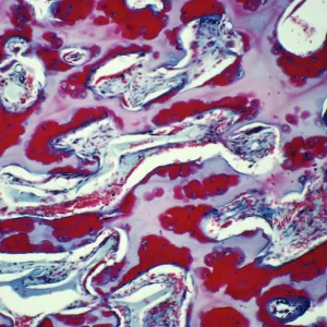
Coloured SEM of femoral spongy bone tissue
Human bone. Coloured scanning electron micrograph of trabeculae in cancellous (spongy) bone tissue of the femur. Bone tissue is divided into compact and cancellous. The latter fills the interior of bones and the cavities between the trabeculae are occupied by bone marrow. Trabeculae are thin and composed of irregular lamellae of bone with lacunae containing osteocytes. Lacunae are visible as tiny, round dark areas on the bone. Osteoblasts are the cells responsible for bone formation. Once they cease activity and become embedded in bone matrix they are called osteocytes. Magnification: x10 at 6x7cm size
Science Photo Library features Science and Medical images including photos and illustrations
Media ID 6448021
© PROF. P. MOTTA/DEPT. OF ANATOMY/UNIVERSITY LA SAPIENZA , ROME/SCIENCE PHOTO LIBRARY
Bones Cancellous Bone Femur Osteocyte Spongy Bone Trabeculae
Photo Mug
Bring the wonders of science into your daily routine with our Media Storehouse Photo Mugs. This unique mug showcases a captivating coloured Scanning Electron Micrograph (SEM) image of femoral spongy bone tissue from Science Photo Library. Delve into the intricacies of human anatomy as you enjoy your favourite beverage. The high-quality print ensures vibrant and long-lasting colours, making each sip an enlightening experience. Perfect for scientists, students, or anyone with a curiosity for the world around us.
A personalised photo mug blends sentimentality with functionality, making an ideal gift for cherished loved ones, close friends, or valued colleagues. Preview may show both sides of the same mug.
Elevate your coffee or tea experience with our premium white ceramic mug. Its wide, comfortable handle makes drinking easy, and you can rely on it to be both microwave and dishwasher safe. Sold in single units, preview may show both sides of the same mug so you can see how the picture wraps around.
Mug Size is 8.1cm high x 9.6cm diameter (3.2" x 3.8")
These are individually made so all sizes are approximate
EDITORS COMMENTS
This print showcases the intricate beauty of femoral spongy bone tissue. In this coloured scanning electron micrograph, we are presented with a mesmerizing view of trabeculae in cancellous (spongy) bone tissue found within the femur. The human bone is divided into two types: compact and cancellous, with the latter filling the interior of bones. The image reveals an array of delicate trabeculae, which are thin structures composed of irregular lamellae of bone. These trabeculae create a network-like pattern that gives strength to the spongy bone tissue. Within each lacuna - tiny dark areas visible on the surface - osteocytes reside as they play their crucial role in maintaining and repairing our bones. Bone marrow fills the cavities between these intricately woven trabeculae, adding further complexity to this microscopic landscape. Osteoblasts, responsible for building new bone material, transform into osteocytes once embedded within the bone matrix. At a magnification level of x10 at 6x7cm size, this photograph allows us to appreciate nature's architectural masterpiece hidden beneath our skin. It serves as a reminder that even at such minuscule scales, there is extraordinary detail and sophistication present in every part of our bodies.
MADE IN AUSTRALIA
Safe Shipping with 30 Day Money Back Guarantee
FREE PERSONALISATION*
We are proud to offer a range of customisation features including Personalised Captions, Color Filters and Picture Zoom Tools
SECURE PAYMENTS
We happily accept a wide range of payment options so you can pay for the things you need in the way that is most convenient for you
* Options may vary by product and licensing agreement. Zoomed Pictures can be adjusted in the Cart.