Photo Mug : Mole nose, SEM


![]()
Home Decor from Science Photo Library
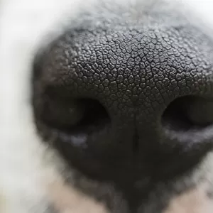
Mole nose, SEM
Mole nose. Coloured scanning electron micrograph (SEM) of the snout (pink) of a mole, showing specialised bulbous papillae (projections) known as Eimers organs. Eimers organs are present in most moles and are made up of epidermal (upper skin layer) cells that are infiltrated by many nerve fibres. These fibres end as a swelling just before the very outer layer of the epidermis, making the snout extremely sensitive to touch. Most moles are virtually blind, so they rely on their specialised ability to sense movement in order to survive
Science Photo Library features Science and Medical images including photos and illustrations
Media ID 6464461
© STEVE GSCHMEISSNER/SCIENCE PHOTO LIBRARY
Bulbous False Colour Hair Hairs Hairy Mammal Mole Nasal Nose Papilla Papillae Sense Sensitive Sensitivity Snout Specialised Tactile Touch False Coloured Specialized
Photo Mug
Add a touch of science to your daily routine with our Media Storehouse Photo Mugs. This unique mug features an intriguing image of a mole's snout, captured in stunning detail through Coloured Scanning Electron Microscopy (SEM) by Science Photo Library. Each sip from this mug brings you closer to the marvels of nature, making your coffee or tea break an educational experience. Perfect for scientists, nature enthusiasts, or anyone with a curious mind, this mug is a must-have addition to your collection. Embrace the beauty of science with every use.
A personalised photo mug blends sentimentality with functionality, making an ideal gift for cherished loved ones, close friends, or valued colleagues. Preview may show both sides of the same mug.
Elevate your coffee or tea experience with our premium white ceramic mug. Its wide, comfortable handle makes drinking easy, and you can rely on it to be both microwave and dishwasher safe. Sold in single units, preview may show both sides of the same mug so you can see how the picture wraps around.
Mug Size is 8.1cm high x 9.6cm diameter (3.2" x 3.8")
These are individually made so all sizes are approximate
EDITORS COMMENTS
This print from Science Photo Library showcases the intricate beauty of a mole's nose. In this coloured scanning electron micrograph (SEM), we are granted a close-up view of the snout, which appears in a delicate shade of pink. What makes this image truly fascinating is the presence of specialized bulbous papillae known as Eimers organs. Eimers organs, found in most moles, consist of epidermal cells that have been infiltrated by numerous nerve fibers. These fibers culminate in swellings just before reaching the outermost layer of the skin, rendering the mole's snout exquisitely sensitive to touch. This heightened tactile ability compensates for their limited vision and enables them to survive by relying on their unique capacity to sense movement. The astonishing detail captured in this photograph allows us to appreciate nature's ingenuity at its finest. The hairy surface texture and intricate arrangement of papillae create an awe-inspiring sight that highlights the complexity and adaptability present within biological systems. As we delve into this mesmerizing image, it becomes evident that there is so much more beneath the surface than meets the eye. It serves as a reminder that even seemingly ordinary creatures possess extraordinary attributes worthy of exploration and admiration.
MADE IN AUSTRALIA
Safe Shipping with 30 Day Money Back Guarantee
FREE PERSONALISATION*
We are proud to offer a range of customisation features including Personalised Captions, Color Filters and Picture Zoom Tools
SECURE PAYMENTS
We happily accept a wide range of payment options so you can pay for the things you need in the way that is most convenient for you
* Options may vary by product and licensing agreement. Zoomed Pictures can be adjusted in the Cart.