Home > Popular Themes > Human Body
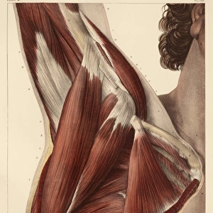
Hand muscle anatomy, 1831 artwork

![]()
Wall Art and Photo Gifts from Science Photo Library
Hand muscle anatomy, 1831 artwork
Hand muscle anatomy. Posterior view of the ligaments and muscles of the hand (main artwork, left). The insets show wrist ligaments (upper right) and a sectioned finger (lower right). This anatomical artwork is plate 122 from volume 2 (1831) of Traite complet de l anatomie de l homme (1831-1854). This 8-volume anatomy atlas was produced by the French physician and anatomist Jean-Baptiste Marc Bourgery (1797-1849). The illustrations were by Nicolas-Henri Jacob (1781-1871)
Science Photo Library features Science and Medical images including photos and illustrations
Media ID 9222663
© SCIENCE PHOTO LIBRARY
1831 Anatomical Artwork Anatomical Illustration Anatomy Atlas Bones Finger Fingers French From Behind Hand Inset Insets Jean Baptiste Marc Bourgery Ligament Ligaments Muscles Nicolas Henri Jacob Phalanges Phalanx Posterior View Volume 2 Volume Ii Wrist Musculature Section Sectioned
MADE IN AUSTRALIA
Safe Shipping with 30 Day Money Back Guarantee
FREE PERSONALISATION*
We are proud to offer a range of customisation features including Personalised Captions, Color Filters and Picture Zoom Tools
SECURE PAYMENTS
We happily accept a wide range of payment options so you can pay for the things you need in the way that is most convenient for you
* Options may vary by product and licensing agreement. Zoomed Pictures can be adjusted in the Cart.