Home > Popular Themes > Human Body
Tooth cross-section, artwork

![]()
Wall Art and Photo Gifts from Science Photo Library
Tooth cross-section, artwork
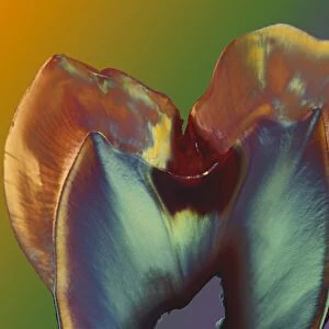
Tooth cross-section, artwork. The upper (biting) surfaces of the tooth are at top, with the lower sections (bottom) embedded in the gums and jaw bone (not shown). The cross-section shows the tooths internal anatomy, including the living tissue of the pulp (red) in the tooths core and its roots (bottom). Within the pulp are venous (blue) and arterial (red) capillaries (small blood vessels), as well as nerves (orange). It is inflammation and infection of this living tissue that causes the pain of a toothache if tooth decay causes loss of the overlying inorganic enamel and dentine layers. This tooth is one of the molars, used for chewing
Science Photo Library features Science and Medical images including photos and illustrations
Media ID 9242983
© CLAUS LUNAU/SCIENCE PHOTO LIBRARY
Chamber Crown Dental Dentin Dentistry Enamel Internal Structure Molar Nerve Nerves Oral Pulp Root Root Canal Roots Tooth Vessels Blood Vessel Cutouts Nervous System
EDITORS COMMENTS
This print showcases a detailed cross-section of a tooth, revealing its intricate internal anatomy. The upper surface of the tooth, used for biting, is displayed at the top, while the lower sections are concealed within the gums and jaw bone. The artwork highlights the living tissue of the pulp in vibrant red coloration, situated at the core of the tooth along with its roots extending downwards. Within this pulp lies a network of venous and arterial capillaries depicted in blue and red respectively, responsible for supplying blood to nourish this vital organ. Additionally, orange nerves can be observed intertwining throughout this complex structure. It is important to note that inflammation or infection affecting this living tissue can lead to excruciating toothaches if decay erodes away the protective enamel and dentine layers above. The featured molar tooth holds significant importance as it plays a crucial role in chewing food effectively. This stunning still life image on a white background provides an educational insight into dental anatomy and serves as a valuable resource for professionals in fields such as biology, medicine, dentistry, and history alike. Claus Lunau's artistic representation beautifully captures both scientific accuracy and aesthetic appeal within this single photograph print from Science Photo Library.
MADE IN AUSTRALIA
Safe Shipping with 30 Day Money Back Guarantee
FREE PERSONALISATION*
We are proud to offer a range of customisation features including Personalised Captions, Color Filters and Picture Zoom Tools
SECURE PAYMENTS
We happily accept a wide range of payment options so you can pay for the things you need in the way that is most convenient for you
* Options may vary by product and licensing agreement. Zoomed Pictures can be adjusted in the Cart.