Canvas Print > Animals > Mammals > Muridae > Fortior
Canvas Print : Removed kneecap, X-ray C017 / 7556


![]()
Canvas Prints from Science Photo Library
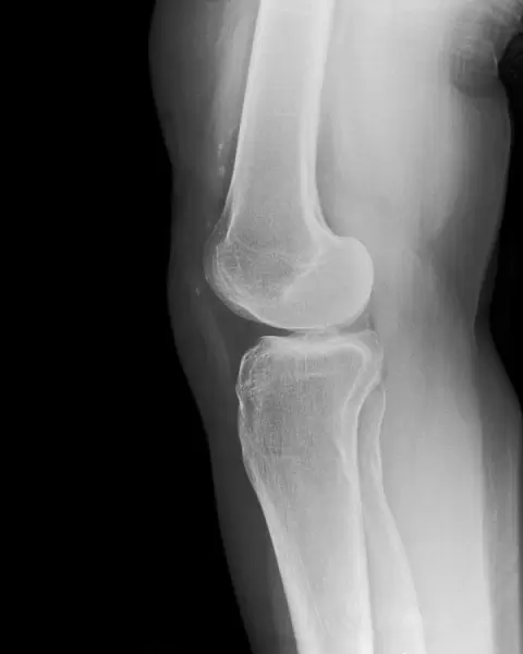
Removed kneecap, X-ray C017 / 7556
Removed kneecap. X-ray of the knee of a 46 year old female that has had her kneecap (patella) surgically removed in a procedure known as a patellectomy. The patella may be removed due to severe injury or disease, such as arthritis
Science Photo Library features Science and Medical images including photos and illustrations
Media ID 9260651
© SCIENCE PHOTO LIBRARY
Forties Forty Six Joint Knee Middle Aged Orthopaedics Orthopedics Patella Patient Profile Radiography Removal Removed X Ray Machine Xray Abnormal Unhealthy
20"x16" (51x41cm) Canvas Print
Discover the intrigue of human anatomy with our Media Storehouse Canvas Prints featuring "Removed kneecap, X-ray C017 / 7556" by Science Photo Library. This captivating image showcases an X-ray of a 46-year-old female's knee post-patellectomy, revealing the absence of her kneecap. These high-quality canvas prints are not only a unique addition to any medical or educational space but also an intriguing conversation starter. Bring the mystery of the human body into your home or office with this striking piece of art.
Delivered stretched and ready to hang our premium quality canvas prints are made from a polyester/cotton blend canvas and stretched over a 1.25" (32mm) kiln dried knot free wood stretcher bar. Packaged in a plastic bag and secured to a cardboard insert for safe transit.
Canvas Prints add colour, depth and texture to any space. Professionally Stretched Canvas over a hidden Wooden Box Frame and Ready to Hang
Estimated Product Size is 40.6cm x 50.8cm (16" x 20")
These are individually made so all sizes are approximate
Artwork printed orientated as per the preview above, with portrait (vertical) orientation to match the source image.
EDITORS COMMENTS
This photo print, titled "Removed kneecap, X-ray C017 / 7556" offers a glimpse into the intricate world of orthopedic medicine. The image showcases the profile view of a 46-year-old female patient who has undergone a patellectomy, a surgical procedure involving the removal of her kneecap (patella). The reasons for such an intervention can vary from severe injury to debilitating diseases like arthritis. As we observe this X-ray, we witness the absence of this vital bone structure in her knee joint. It serves as a stark reminder of the challenges faced by individuals battling with unhealthy joints. The monochrome radiography highlights not only the abnormality but also emphasizes the resilience and adaptability of our bodies. This middle-aged woman's determination to seek medical intervention is evident through this powerful visual representation. Through Science Photo Library's lens, we gain insight into both the fragility and strength inherent within our skeletal system. This thought-provoking image prompts us to reflect on how modern medicine can alleviate pain and improve quality of life for those suffering from joint-related conditions.
MADE IN AUSTRALIA
Safe Shipping with 30 Day Money Back Guarantee
FREE PERSONALISATION*
We are proud to offer a range of customisation features including Personalised Captions, Color Filters and Picture Zoom Tools
SECURE PAYMENTS
We happily accept a wide range of payment options so you can pay for the things you need in the way that is most convenient for you
* Options may vary by product and licensing agreement. Zoomed Pictures can be adjusted in the Cart.