
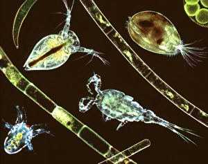
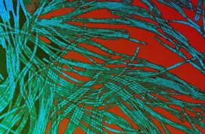
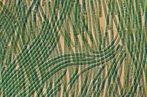
Filamentous Collection
"Exploring the Intricate World Cell Structures in Basidiomycota: A Fascinating Journey into the Microscopic Realm" In the realm of biology


All Professionally Made to Order for Quick Shipping
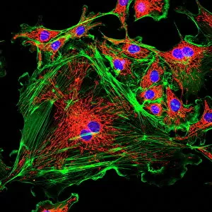
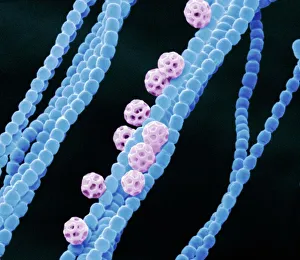
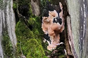
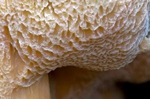
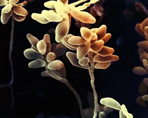
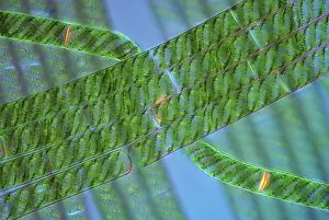
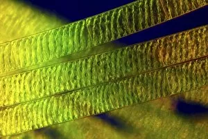
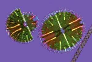
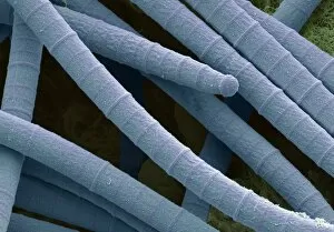
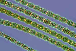
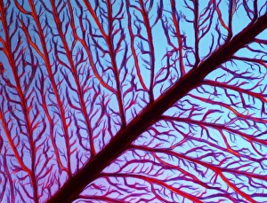
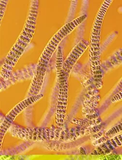
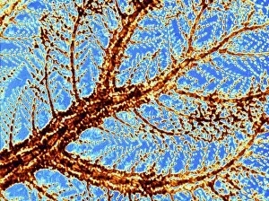
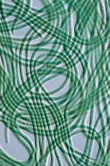
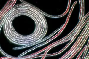
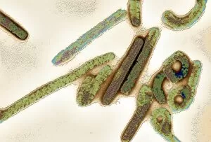
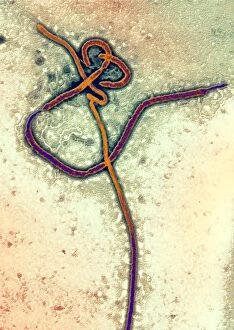
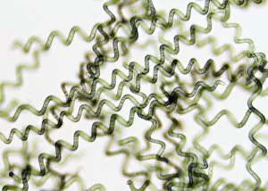
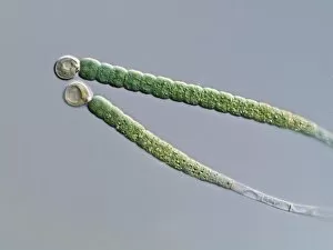
"Exploring the Intricate World Cell Structures in Basidiomycota: A Fascinating Journey into the Microscopic Realm" In the realm of biology, filamentous structures hold a significant place, particularly in the study of fungi. Basidiomycota, a diverse group fungi, captivate scientists with their intricate cellular arrangements. These organisms exhibit an array of fascinating characteristics that are worth delving into. One captivating aspect is their association with cyanobacteria. Through scanning electron microscopy (SEM), researchers have uncovered symbiotic relationships between these two entities. The images captured through this technique showcase the delicate intertwining filaments and provide insights into how these organisms interact at a microscopic level. Amongst the vast collection of images depicting cell structures, BAL0651801 stands out as it offers an exquisite glimpse into the world fungi. This particular image showcases brown-rot Postia placenta's close-up detail of pores found in Clumber Park, Nottinghamshire, England during October. The intricate network formed by these filaments highlights their role in decomposition processes and nutrient cycling within ecosystems. As we explore further through Picture No. 11675027 to Picture No. 10890444, we witness various other mesmerizing examples showcasing different aspects and species within this unique group. Each image tells its own story about filamentous structures - from their diversity to their ecological significance. These captivating visuals not only serve as scientific documentation but also ignite curiosity among those who appreciate nature's hidden wonders on a microscopic scale. They remind us that even within seemingly ordinary environments like forests or parks lie extraordinary intricacies waiting to be discovered. So let us embark on this journey together – unraveling the mysteries held within each filamentous structure – for there is much more than meets the eye when it comes to understanding life's smallest building blocks.