Framed Print : Frog embryo, light micrograph


![]()
Framed Photos from Science Photo Library
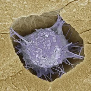
Frog embryo, light micrograph
Frog embryo. Light micrograph of a frog embryo at the blastula stage. This stage in the embryos development is produced by the cleavage (cell division) of a fertilised ovum (egg cell). At this point the embryo is a hollow ball of cells surrounding a central fluid-filled cavity called the blastocoel. The outer layer of cells is known as the blastoderm. At bottom is the vegetal pole, which will develop into the endoderm. The endoderm forms the digestive tract, glands and the lungs. Magnification: x30 when printed at 10 centimetres wide
Science Photo Library features Science and Medical images including photos and illustrations
Media ID 6455567
© STEVE GSCHMEISSNER/SCIENCE PHOTO LIBRARY
Developing Development Developmental Biology Embryo Embryology Endoderm Frog Re Production Reproductive Stage Blastocoel Blastula Cells Light Micrograph Light Microscope
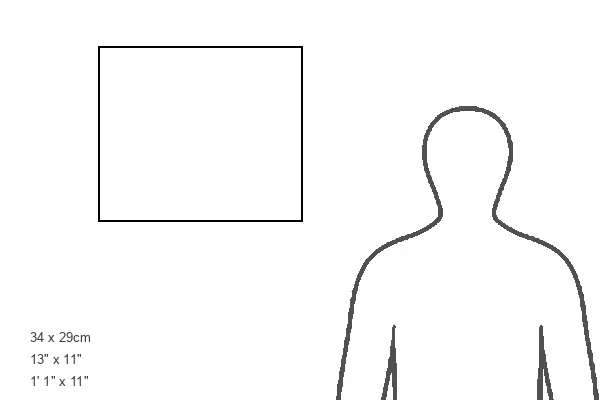
13.5"x11.5" (34x29cm) Premium Frame
Discover the wonders of nature with Media Storehouse's Framed Prints featuring the fascinating image of a frog embryo at the blastula stage. This captivating light micrograph from Science Photo Library showcases the intricate process of cell division during the early stages of an embryo's development. Bring this mesmerizing piece of science into your home or office as a conversation starter and a reminder of the beauty and complexity of life.
Framed and mounted 9x7 print. Professionally handmade full timber moulded frames are finished off with framers tape and come with a hanging solution on the back. Outer dimensions are 13.5x11.5 inches (34x29cm). Quality timber frame frame moulding (20mm wide and 30mm deep) with frame colours in your choice of black, white, or raw oak and a choice of black or white card mounts. Frames have a perspex front providing a virtually unbreakable glass-like finish which is easily cleaned with a damp cloth.
Contemporary Framed and Mounted Prints - Professionally Made and Ready to Hang
Estimated Image Size (if not cropped) is 21.4cm x 21.4cm (8.4" x 8.4")
Estimated Product Size is 34cm x 29.2cm (13.4" x 11.5")
These are individually made so all sizes are approximate
Artwork printed orientated as per the preview above, with landscape (horizontal) or portrait (vertical) orientation to match the source image.
EDITORS COMMENTS
This print captures the intricate beauty of a frog embryo at the blastula stage. The image, taken using a light microscope, showcases the remarkable process of cell division and development in these tiny creatures. At this stage, which is produced by the cleavage of a fertilized egg cell, the embryo takes on the form of a hollow ball composed of cells surrounding a central fluid-filled cavity known as the blastocoel. The outer layer of cells is referred to as the blastoderm. In this mesmerizing micrograph, we can observe various key features. At the bottom lies the vegetal pole, which will eventually develop into an essential part called endoderm. This vital layer gives rise to crucial structures such as digestive tracts, glands, and even lungs. The magnification used for this print allows us to appreciate every intricate detail present in this developing organism. With each passing moment during embryonic development, nature's wonders unfold before our eyes. This photograph serves not only as an awe-inspiring piece showcasing nature's incredible ability to create life but also provides valuable insights into developmental biology and reproductive processes. It reminds us that within every single living being lies an extraordinary journey from conception to birth—a testament to both science and artistry captured by Science Photo Library.
MADE IN AUSTRALIA
Safe Shipping with 30 Day Money Back Guarantee
FREE PERSONALISATION*
We are proud to offer a range of customisation features including Personalised Captions, Color Filters and Picture Zoom Tools
SECURE PAYMENTS
We happily accept a wide range of payment options so you can pay for the things you need in the way that is most convenient for you
* Options may vary by product and licensing agreement. Zoomed Pictures can be adjusted in the Cart.