Fraud Blocker Skip to main content

Anatomist Collection (page 4)

Anatomists have played a crucial role in unraveling the mysteries of the human body throughout history

Background imageAnatomist Collection: Surgical instruments for extracting foreign bodies from the esophagus - Plate from '

Surgical instruments for extracting foreign bodies from the esophagus - Plate from " L'Encyclopedie" by
FLO4611943 Surgical instruments for extracting foreign bodies from the esophagus - Plate from " L'Encyclopedie" by Denis Diderot (1713-1784) and Jean Le Rond D'Alembert (1717-1783)

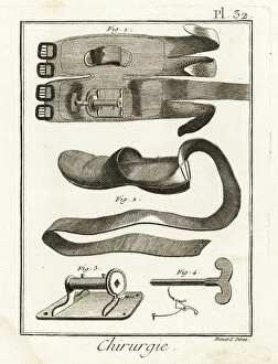
Background imageAnatomist Collection: Knee pad, slipper and mechanical mechanism - Plate from ' L'Encyclopedie' by Denis Diderot

Knee pad, slipper and mechanical mechanism - Plate from " L'Encyclopedie" by Denis Diderot (1713-1784)
FLO4611907 Knee pad, slipper and mechanical mechanism - Plate from " L'Encyclopedie" by Denis Diderot (1713-1784) and Jean Le Rond D'Alembert (1717-1783)

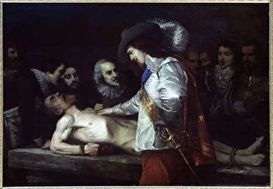
Background imageAnatomist Collection: The british physiologist William Harvey and the systemic circulation

The british physiologist William Harvey and the systemic circulation. Painting, France, 18th century (oil on canvas)
3434349 The british physiologist William Harvey and the systemic circulation. Painting, France, 18th century (oil on canvas); Academie des Sciences, Paris, France; Photo © CCI.

Background imageAnatomist Collection: Shi si jing fa hui

Shi si jing fa hui
2626560 Shi si jing fa hui; (add.info.: Illustration from the Japanese & Chinese edition (1716) of 'Shi si jing fa hui'

Background imageAnatomist Collection: Shi si jing fa hui

Shi si jing fa hui
2626556 Shi si jing fa hui; (add.info.: Illustration from the Japanese & Chinese edition (1716) of 'Shi si jing fa hui'

Background imageAnatomist Collection: Fasciculus medicinae

Fasciculus medicinae
2626472 Fasciculus medicinae; (add.info.: Illustration from 'Fasciculus medicinae' Venice; 1494 the first printed book to contain anatomical illustrations)

Background imageAnatomist Collection: The Anatomy of Human Bodies, 1698 (engraving)

The Anatomy of Human Bodies, 1698 (engraving)
2625659 The Anatomy of Human Bodies, 1698 (engraving); (add.info.: Illustration from 'The anatomy of humane bodies' by William Cowper, (Oxford, 1698).); Universal History Archive/UIG.

Background imageAnatomist Collection: Synoptic Chart of Anatomy

Synoptic Chart of Anatomy
2625362 Synoptic Chart of Anatomy; (add.info.: Synoptic charts concerning the anatomy of the upper and lower jaw (fakk), from the second chapter (bab) of the second book (kit)

Background imageAnatomist Collection: Equine anatomical Illustration

Equine anatomical Illustration
2626343 Equine anatomical Illustration; (add.info.: Equine anatomical Illustration from 'Anatomia del cavallo, infermita, et suoi rimedii')

Background imageAnatomist Collection: Equine anatomical Illustration

Equine anatomical Illustration
2626313 Equine anatomical Illustration; (add.info.: Equine anatomical Illustration from 'Anatomia del cavallo, infermita, et suoi rimedii')

Background imageAnatomist Collection: Equine anatomical Illustration

Equine anatomical Illustration
2626310 Equine anatomical Illustration; (add.info.: Equine anatomical Illustration from 'Anatomia del cavallo, infermita, et suoi rimedii')

Background imageAnatomist Collection: Shi si jing fa hui

Shi si jing fa hui
2626541 Shi si jing fa hui; (add.info.: Illustration from the Japanese & Chinese edition (1716) of 'Shi si jing fa hui'

Background imageAnatomist Collection: Fasciculus medicinae

Fasciculus medicinae
2626481 Fasciculus medicinae; (add.info.: Illustration from 'Fasciculus medicinae' Venice; 1494 the first printed book to contain anatomical illustrations)

Background imageAnatomist Collection: Les oeuures d'Ambroise Pare?

Les oeuures d'Ambroise Pare?
2626429 Les oeuures d'Ambroise Pare?; (add.info.: Illustration from 'Les oeuures d'Ambroise Pare?' 1585 by Ambroise Pare)

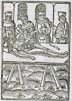
Background imageAnatomist Collection: A dissection in progress. From Le livre de la propriete des choses (On the Properites of Things)

A dissection in progress. From Le livre de la propriete des choses (On the Properites of Things)
527491 A dissection in progress. From Le livre de la propriete des choses (On the Properites of Things) written by the English Franciscan monk Bartholomew Glanville called Bartolomaeus Anglicus

Background imageAnatomist Collection: Anchor escapement mechanism (drawing)

Anchor escapement mechanism (drawing)
535351 Anchor escapement mechanism (drawing); (add.info.: The anchor escapement, which was invented in the 17th century, allowed pendulum clocks to be regulated.); Encyclopaedia Britannica/UIG.

Background imageAnatomist Collection: Johannes Evangelista Purkinje, 1840

Johannes Evangelista Purkinje, 1840
1576997 Johannes Evangelista Purkinje, 1840; (add.info.: Johannes Evangelista Purkinje (17 December or 18 December 1787 - 28 July 1869) was a Czech anatomist and physiologist

Background imageAnatomist Collection: Jacob Henle, 1850

Jacob Henle, 1850
1576988 Jacob Henle, 1850; (add.info.: Jacob Henle, (9 July 1809 - 13 May 1885) was a German physician, pathologist, and anatomist

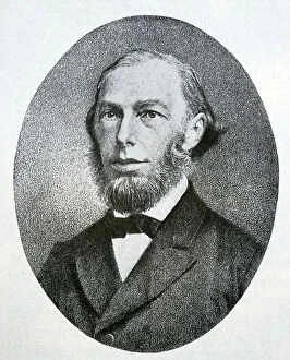
Background imageAnatomist Collection: Sir Richard Owen, 1865

Sir Richard Owen, 1865
1576998 Sir Richard Owen, 1865; (add.info.: Sir Richard Owen (20 July 1804 - 18 December 1892) was an English biologist, comparative anatomist and paleontologist

Background imageAnatomist Collection: Max Johann Sigismund Schultze, 1850

Max Johann Sigismund Schultze, 1850
1576982 Max Johann Sigismund Schultze, 1850; (add.info.: Max Johann Sigismund Schultze (March 25, 1825 - January 16, 1874) was a German microscopic anatomist

Background imageAnatomist Collection: An open female cuttlefish, plate from 'Histoire naturelle: les mollusques' by Georges Louis Leclerc

An open female cuttlefish, plate from "Histoire naturelle: les mollusques" by Georges Louis Leclerc
AIS5369968 An open female cuttlefish, plate from "Histoire naturelle: les mollusques" by Georges Louis Leclerc, comte de Buffon (1707-1788) (engraving); Biblioteca de Catalunya, Barcelona

Background imageAnatomist Collection: Doctor Pibrac's tongue wound bandage and lithotoma, used to hunt the bladder - Plate taken from '

Doctor Pibrac's tongue wound bandage and lithotoma, used to hunt the bladder - Plate taken from "
FLO4611922 Doctor Pibrac's tongue wound bandage and lithotoma, used to hunt the bladder - Plate taken from " L'Encyclopedie" by Denis Diderot (1713-1784)

Background imageAnatomist Collection: Cow tongue under the microscope: veal tongue showing different papillae 1, foliate papilla 2

Cow tongue under the microscope: veal tongue showing different papillae 1, foliate papilla 2, filiform papillae 3
FLO4574832 Cow tongue under the microscope: veal tongue showing different papillae 1, foliate papilla 2, filiform papillae 3, and fungiform papillae 4

Background imageAnatomist Collection: Anatomy of the human nose, mouth, tongue, jaw and teeth, showing the senses of taste

Anatomy of the human nose, mouth, tongue, jaw and teeth, showing the senses of taste, smell and touch
FLO4684860 Anatomy of the human nose, mouth, tongue, jaw and teeth, showing the senses of taste, smell and touch. Lithograph from Lorenz Oken's Universal Natural History

Background imageAnatomist Collection: Scalp diseases, porrigo varietes and skin diseases: ulcerous pyodermatitis caused by bacteria

Scalp diseases, porrigo varietes and skin diseases: ulcerous pyodermatitis caused by bacteria - Lithography by John
FLO4618780 Scalp diseases, porrigo varietes and skin diseases: ulcerous pyodermatitis caused by bacteria - Lithography by John Pass, published in "Encyclopedia Londinensis" by John Wilkes

Background imageAnatomist Collection: Screws for compression of blood vessels by Jean Louis Petite

Screws for compression of blood vessels by Jean Louis Petite, tourniquet device and double band to adjust it
FLO4611834 Screws for compression of blood vessels by Jean Louis Petite, tourniquet device and double band to adjust it, chamois skin compress

Background imageAnatomist Collection: surgical chair with patient to remove a vesical calculation through the groin - Plate taken from '

surgical chair with patient to remove a vesical calculation through the groin - Plate taken from "
FLO4611780 surgical chair with patient to remove a vesical calculation through the groin - Plate taken from " L'Encyclopedie" by Denis Diderot (1713-1784)

Background imageAnatomist Collection: Cranium 1-4 and part of sternum 5,6 of the extinct Rodrigues solitaire

Cranium 1-4 and part of sternum 5,6 of the extinct Rodrigues solitaire. Lithograph by Joseph Dinkel after Werner
FLO4612182 Cranium 1-4 and part of sternum 5,6 of the extinct Rodrigues solitaire. Lithograph by Joseph Dinkel after Werner from Hugh Edwin Strickland and Alexander Gordon Melville's The Dodo

Background imageAnatomist Collection: Skulls of tooth-billed pigeon, Didunculus strigirostris, critically endangered 1,7,8,9,10 dodo

Skulls of tooth-billed pigeon, Didunculus strigirostris, critically endangered 1,7,8,9,10 dodo, Raphus cucullatus 2
FLO4612171 Skulls of tooth-billed pigeon, Didunculus strigirostris, critically endangered 1,7,8,9,10 dodo, Raphus cucullatus 2, yellow-footed green pigeon, Treron chlorigaster 3,6, crowned pigeon

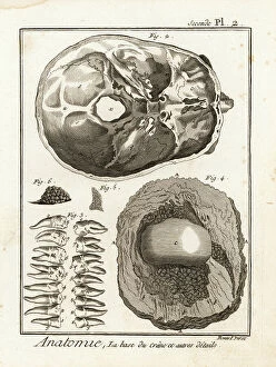
Background imageAnatomist Collection: Base of the skull, human teeth, knee cap (after Clopton Havers), mucus gland, etc

Base of the skull, human teeth, knee cap (after Clopton Havers), mucus gland, etc
FLO4611538 Base of the skull, human teeth, knee cap (after Clopton Havers), mucus gland, etc. Copperplate engraving by Robert Benard from Denis Diderot's Encyclopedia, Pellet, Geneva

Background imageAnatomist Collection: Interior of the brain and cerebellum. Copperplate engraving by Robert Benard after an illustration

Interior of the brain and cerebellum. Copperplate engraving by Robert Benard after an illustration by Albrecht von
FLO4611652 Interior of the brain and cerebellum. Copperplate engraving by Robert Benard after an illustration by Albrecht von Haller from Denis Diderot's Encyclopedia, Pellet, Geneva

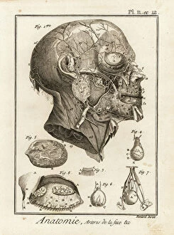
Background imageAnatomist Collection: Arteries in the face. Copperplate engraving by Robert Benard after an illustration by Albrecht von

Arteries in the face. Copperplate engraving by Robert Benard after an illustration by Albrecht von Haller from Denis
FLO4611618 Arteries in the face. Copperplate engraving by Robert Benard after an illustration by Albrecht von Haller from Denis Diderot's Encyclopedia, Pellet, Geneva

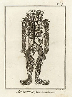
Background imageAnatomist Collection: Venal system from the vena cava in the human body. Copperplate engraving by Robert Benard after an

Venal system from the vena cava in the human body. Copperplate engraving by Robert Benard after an illustration by
FLO4611599 Venal system from the vena cava in the human body. Copperplate engraving by Robert Benard after an illustration by James Drake from Denis Diderot's Encyclopedia, Pellet, Geneva

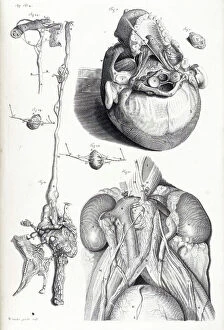
Background imageAnatomist Collection: Details of arteries, veins, lymph acorns, etc. Copperplate engraving by Robert Benard after

Details of arteries, veins, lymph acorns, etc. Copperplate engraving by Robert Benard after from Denis Diderot's
FLO4611594 Details of arteries, veins, lymph acorns, etc. Copperplate engraving by Robert Benard after from Denis Diderot's Encyclopedia, Pellet, Geneva, 1779.; (add.info.: Details of arteries)

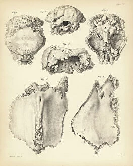
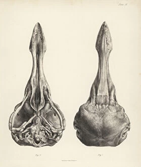
Background imageAnatomist Collection: Upper and lower views of the skull of a dodo. Lithograph from Hugh Edwin Strickland

Upper and lower views of the skull of a dodo. Lithograph from Hugh Edwin Strickland
FLO4612153 Upper and lower views of the skull of a dodo. Lithograph from Hugh Edwin Strickland and Alexander Gordon Melville's The Dodo and its Kindred, London, Reeve, Benham and Reeve

Background imageAnatomist Collection: Tooth billed pigeon, Didunculus strigirostris 1,2 (critically endangered), Bruce's green pigeon

Tooth billed pigeon, Didunculus strigirostris 1,2 (critically endangered), Bruce's green pigeon
FLO4612139 Tooth billed pigeon, Didunculus strigirostris 1,2 (critically endangered), Bruce's green pigeon, Treron waalia (Treron abyssinica 3,5) and Levaillant's dove

Background imageAnatomist Collection: Jean Louis Petite's machine to compress femoral artere during thigh amputation, bandage

Jean Louis Petite's machine to compress femoral artere during thigh amputation, bandage
FLO4611844 Jean Louis Petite's machine to compress femoral artere during thigh amputation, bandage, aneurysm needle and scissors - Plate taken from " L'Encyclopedie" by Denis

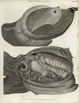
Background imageAnatomist Collection: Anatomy of the horse: stomach and intestines. Copperplate engraving by Scott from Abraham Rees'

Anatomy of the horse: stomach and intestines. Copperplate engraving by Scott from Abraham Rees' Cyclopedia or
FLO4593968 Anatomy of the horse: stomach and intestines. Copperplate engraving by Scott from Abraham Rees' Cyclopedia or Universal Dictionary of Arts, Sciences and Literature, Longman, Hurst

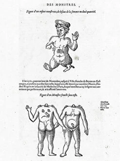
Background imageAnatomist Collection: Engraving of the book of Ambroise Pare: ' Monsters and Prodiges' (first edition 1573)

Engraving of the book of Ambroise Pare: " Monsters and Prodiges" (first edition 1573)
PCT4260964 Engraving of the book of Ambroise Pare: " Monsters and Prodiges" (first edition 1573, appendix: marine monsters)

Background imageAnatomist Collection: Le grand sperm whale in ' Painting Encyclopedique et Methodique des trois Regnes de la Nature' by

Le grand sperm whale in " Painting Encyclopedique et Methodique des trois Regnes de la Nature" by Pierre
FAT4299958 Le grand sperm whale in " Painting Encyclopedique et Methodique des trois Regnes de la Nature" by Pierre Joseph Bonnaterre (1762-1804), Paris

Background imageAnatomist Collection: Skeleton of a European perch, Perca fluviatilis, from Dr. Barclay's Museum, Edinburgh

Skeleton of a European perch, Perca fluviatilis, from Dr. Barclay's Museum, Edinburgh
FLO4565818 Skeleton of a European perch, Perca fluviatilis, from Dr. Barclay's Museum, Edinburgh. Copperplate engraving by Edward Mitchell after an anatomical illustration by Robert Kaye Greville

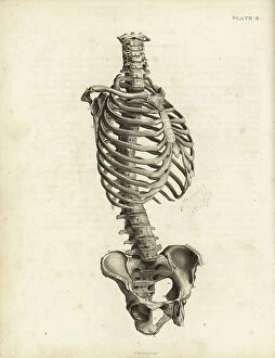
Background imageAnatomist Collection: Rear view of the human trunk skeleton including spine, ribs and pelvis

Rear view of the human trunk skeleton including spine, ribs and pelvis. Copperplate engraving by Edward Mitchell after
FLO4565792 Rear view of the human trunk skeleton including spine, ribs and pelvis. Copperplate engraving by Edward Mitchell after an anatomical illustration by Jean-Joseph Sue from John Barclay's

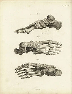
Background imageAnatomist Collection: Views of the bones in the human foot from ankle to toe. Copperplate engraving by Edward Mitchell

Views of the bones in the human foot from ankle to toe. Copperplate engraving by Edward Mitchell after an anatomical
FLO4565738 Views of the bones in the human foot from ankle to toe. Copperplate engraving by Edward Mitchell after an anatomical illustration by Jean-Joseph Sue from John Barclay's A Series of

Background imageAnatomist Collection: Bones of the carpus, metacarpus and fingers in the human hand

Bones of the carpus, metacarpus and fingers in the human hand. Copperplate engraving by Edward Mitchell after an
FLO4565722 Bones of the carpus, metacarpus and fingers in the human hand. Copperplate engraving by Edward Mitchell after an anatomical illustration from John Barclay's A Series of Engravings of

Background imageAnatomist Collection: Sternum and cartilage of the human ribcage, with several ribs

Sternum and cartilage of the human ribcage, with several ribs. Copperplate engraving by Edward Mitchell after an
FLO4565702 Sternum and cartilage of the human ribcage, with several ribs. Copperplate engraving by Edward Mitchell after an anatomical illustration by Jean-Joseph Sue from John Barclay's A Series

Background imageAnatomist Collection: Ribs and sternum in the human ribcage. Copperplate engraving by Edward Mitchell after an

Ribs and sternum in the human ribcage. Copperplate engraving by Edward Mitchell after an anatomical illustration by
FLO4565699 Ribs and sternum in the human ribcage. Copperplate engraving by Edward Mitchell after an anatomical illustration by Jean-Joseph Sue from John Barclay's A Series of Engravings of

Background imageAnatomist Collection: Front view of the human trunk skeleton including spine, ribs and pelvis

Front view of the human trunk skeleton including spine, ribs and pelvis. Copperplate engraving by Edward Mitchell after
FLO4565690 Front view of the human trunk skeleton including spine, ribs and pelvis. Copperplate engraving by Edward Mitchell after an anatomical illustration by Jean-Joseph Suefrom John Barclay's

Background imageAnatomist Collection: Prickly cockle - bucarde - Acanthocardia echinata (Cardium echinatum) 1

Prickly cockle - bucarde - Acanthocardia echinata (Cardium echinatum) 1, and slender sea pen - plume elegante
FLO4576265 Prickly cockle - bucarde - Acanthocardia echinata (Cardium echinatum) 1, and slender sea pen - plume elegante - Virgularia mirabilis (Pennatula mirabilis) 2


All Professionally Made to Order for Quick Shipping

Anatomists have played a crucial role in unraveling the mysteries of the human body throughout history. One such pioneer was Robert Burton, whose extensive research and writings on anatomy left an indelible mark on the field. Another notable figure was EUSTACHIO Bartolomeo, an Italian anatomist who made significant contributions during the 16th century. Intriguingly, a lithograph titled "Odin questioning Mimir" captures the essence of curiosity that drives anatomists to seek knowledge. This thirst for understanding is exemplified by Sir Richard Owen, a renowned British anatomist from the 19th century whose work continues to inspire modern scientists. Richard Owen's dedication to his craft is evident in various depictions of him throughout history. In one caricature from Vanity Fair in 1873, aptly titled "Old bones, " he is portrayed as a distinguished scholar with wisdom etched onto his face. An image from his study at BMNH in 1883 showcases his meticulous approach towards dissecting and studying specimens. The intricate details captured in engravings like "The Sacred Heart of Jesus" or "The pollen of plants" demonstrate how anatomists explore not only human anatomy but also delve into other aspects of nature's design. Richard Owen himself had a profound impact on paleontology, as depicted in C016 / 5008 - an image that pays homage to his groundbreaking discoveries. Throughout history, heart anatomists have held a special place within this scientific realm. Figures like Guillaume Dupuytren and William Hunter dedicated their lives to unraveling the complexities of this vital organ. Their contributions paved the way for advancements in cardiac medicine that continue to save lives today. From ancient times until now, these brilliant minds have shaped our understanding of ourselves and our world through their tireless pursuit of knowledge about our bodies' inner workings. The legacy they leave behind serves as an inspiration for future generations aspiring to unravel the secrets of human anatomy.