Fraud Blocker Skip to main content

Anatomist Collection (page 5)

Anatomists have played a crucial role in unraveling the mysteries of the human body throughout history

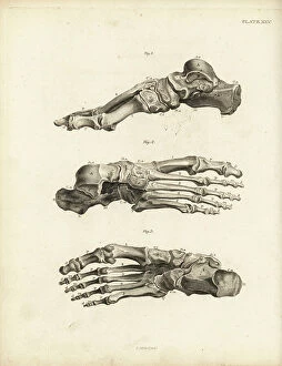
Background imageAnatomist Collection: Views of the bones in the human foot from ankle to toe. Copperplate engraving by Edward Mitchell

Views of the bones in the human foot from ankle to toe. Copperplate engraving by Edward Mitchell after an anatomical
FLO4565738 Views of the bones in the human foot from ankle to toe. Copperplate engraving by Edward Mitchell after an anatomical illustration by Jean-Joseph Sue from John Barclay's A Series of

Background imageAnatomist Collection: Bones of the carpus, metacarpus and fingers in the human hand

Bones of the carpus, metacarpus and fingers in the human hand. Copperplate engraving by Edward Mitchell after an
FLO4565722 Bones of the carpus, metacarpus and fingers in the human hand. Copperplate engraving by Edward Mitchell after an anatomical illustration from John Barclay's A Series of Engravings of

Background imageAnatomist Collection: Sternum and cartilage of the human ribcage, with several ribs

Sternum and cartilage of the human ribcage, with several ribs. Copperplate engraving by Edward Mitchell after an
FLO4565702 Sternum and cartilage of the human ribcage, with several ribs. Copperplate engraving by Edward Mitchell after an anatomical illustration by Jean-Joseph Sue from John Barclay's A Series

Background imageAnatomist Collection: Ribs and sternum in the human ribcage. Copperplate engraving by Edward Mitchell after an

Ribs and sternum in the human ribcage. Copperplate engraving by Edward Mitchell after an anatomical illustration by
FLO4565699 Ribs and sternum in the human ribcage. Copperplate engraving by Edward Mitchell after an anatomical illustration by Jean-Joseph Sue from John Barclay's A Series of Engravings of

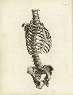
Background imageAnatomist Collection: Front view of the human trunk skeleton including spine, ribs and pelvis

Front view of the human trunk skeleton including spine, ribs and pelvis. Copperplate engraving by Edward Mitchell after
FLO4565690 Front view of the human trunk skeleton including spine, ribs and pelvis. Copperplate engraving by Edward Mitchell after an anatomical illustration by Jean-Joseph Suefrom John Barclay's

Background imageAnatomist Collection: Prickly cockle - bucarde - Acanthocardia echinata (Cardium echinatum) 1

Prickly cockle - bucarde - Acanthocardia echinata (Cardium echinatum) 1, and slender sea pen - plume elegante
FLO4576265 Prickly cockle - bucarde - Acanthocardia echinata (Cardium echinatum) 1, and slender sea pen - plume elegante - Virgularia mirabilis (Pennatula mirabilis) 2

Background imageAnatomist Collection: Adult crane desarticule 1867

Adult crane desarticule 1867
JAB4120023 Adult crane desarticule 1867; (add.info.: Adult crane desarticule 1867); Photo ©Jaime Abecasis.

Background imageAnatomist Collection: Bladder anatomy 1866 illustrations showing the internal (top) and external (bottom)

Bladder anatomy 1866 illustrations showing the internal (top) and external (bottom)
JAB4119242 Bladder anatomy 1866 illustrations showing the internal (top) and external (bottom) anatomy of the male urinary bladder, showing its muscular coating (brown)

Background imageAnatomist Collection: Voice box and thyroid 1866 illustration showing the anatomy of the larynx (voice box)

Voice box and thyroid 1866 illustration showing the anatomy of the larynx (voice box) and thyroid (brown)
JAB4119213 Voice box and thyroid 1866 illustration showing the anatomy of the larynx (voice box) and thyroid (brown, lower centre) Shown here are the hyoid bone (A), thyroid cartilage (B)

Background imageAnatomist Collection: Chest muscles - 1866 illustration showing the structure of the muscles (brown) and tendons (white)

Chest muscles - 1866 illustration showing the structure of the muscles (brown) and tendons (white) of the human chest
JAB4119479 Chest muscles - 1866 illustration showing the structure of the muscles (brown) and tendons (white) of the human chest

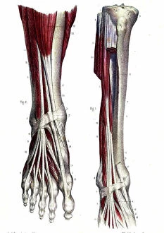
Background imageAnatomist Collection: Leg and foot muscles - 1866 anatomical illustration showing the muscles (brown) and tendons (white)

Leg and foot muscles - 1866 anatomical illustration showing the muscles (brown) and tendons (white)
JAB4119476 Leg and foot muscles - 1866 anatomical illustration showing the muscles (brown) and tendons (white) of the right foot (left)

Background imageAnatomist Collection: Shoulder muscles - 1866 illustration of the muscles (brown) and tendons (white)

Shoulder muscles - 1866 illustration of the muscles (brown) and tendons (white)
JAB4119462 Shoulder muscles - 1866 illustration of the muscles (brown) and tendons (white) associated with the human collarbone (clavicle, top) and shoulder blade (scapula)

Background imageAnatomist Collection: Chest and shoulder muscles - 1866 anatomical illustration showing the structure of the muscles

Chest and shoulder muscles - 1866 anatomical illustration showing the structure of the muscles (brown)
JAB4119433 Chest and shoulder muscles - 1866 anatomical illustration showing the structure of the muscles (brown) and tendons (white)

Background imageAnatomist Collection: Hand muscles - 1866 anatomical illustration showing the muscles (brown) and tendons (white)

Hand muscles - 1866 anatomical illustration showing the muscles (brown) and tendons (white) of the human hand
JAB4119426 Hand muscles - 1866 anatomical illustration showing the muscles (brown) and tendons (white) of the human hand, with insets (left)

Background imageAnatomist Collection: Rib cage anatomy - 1866 illustration showing the bones of the human rib cage

Rib cage anatomy - 1866 illustration showing the bones of the human rib cage, including the ribs (bottom)
JAB4119411 Rib cage anatomy - 1866 illustration showing the bones of the human rib cage, including the ribs (bottom), spine (centre-left) and sternum (breast bone, centre and upper right) Le Thorax

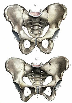
Background imageAnatomist Collection: Pelvis anatomy - 1866 illustration showing the anatomy of the male (top) and female (bottom)

Pelvis anatomy - 1866 illustration showing the anatomy of the male (top) and female (bottom) pelvis - The pelvis
JAB4119404 Pelvis anatomy - 1866 illustration showing the anatomy of the male (top) and female (bottom) pelvis - The pelvis, anatomy, osteology - Fig 1: female pelvis Fig 2: male pelvis, Musculotion

Background imageAnatomist Collection: Hip muscles - 1866 illustration showing the structure of the muscles (brown) and tendons (white)

Hip muscles - 1866 illustration showing the structure of the muscles (brown) and tendons (white)
JAB4119401 Hip muscles - 1866 illustration showing the structure of the muscles (brown) and tendons (white) of the upper right leg and hip

Background imageAnatomist Collection: Arm muscles - 1866 illustration showing the structure of the muscles (brown) and tendons (white)

Arm muscles - 1866 illustration showing the structure of the muscles (brown) and tendons (white)
JAB4119391 Arm muscles - 1866 illustration showing the structure of the muscles (brown) and tendons (white) of the human arm and top of the hand (bottom left and right)

Background imageAnatomist Collection: Abdominal muscles - 1866 illustration showing the structure of the muscles (brown)

Abdominal muscles - 1866 illustration showing the structure of the muscles (brown) and tendons (white)
JAB4119387 Abdominal muscles - 1866 illustration showing the structure of the muscles (brown) and tendons (white) found in the rear of the human trunk - L'abdomen - Anatomy - Region anterieure de

Background imageAnatomist Collection: Leg muscles - 1866 illustration showing the structure of the muscles (brown) and tendons (white)

Leg muscles - 1866 illustration showing the structure of the muscles (brown) and tendons (white)
JAB4119363 Leg muscles - 1866 illustration showing the structure of the muscles (brown) and tendons (white) of the rear of the lower leg - Anatomy du dos de la leg - Fig 1 et 2

Background imageAnatomist Collection: Andreas Vesalius

Andreas Vesalius
3586025 Andreas Vesalius; (add.info.: Portrait of Andreas Vesalius (1514-1564) a Flemish/Netherlandish anatomist, physician, and author of books on human anatomy

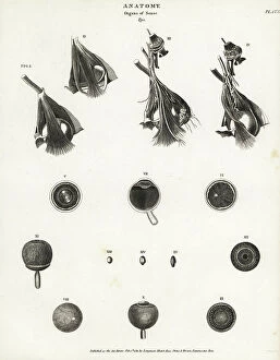
Background imageAnatomist Collection: Anatomy of the human eye, showing eyeball, optic nerve, muscle, cornea, retina, sections through eye

Anatomy of the human eye, showing eyeball, optic nerve, muscle, cornea, retina, sections through eye
FLO4591961 Anatomy of the human eye, showing eyeball, optic nerve, muscle, cornea, retina, sections through eye. Copperplate engraving by Milton from Abraham Rees' Cyclopedia or Universal

Background imageAnatomist Collection: Anatomy of human internal organs from the back showing stomach, liver, intestines, gallbladder, etc

Anatomy of human internal organs from the back showing stomach, liver, intestines, gallbladder, etc
FLO4590759 Anatomy of human internal organs from the back showing stomach, liver, intestines, gallbladder, etc. Copperplate engraving by Milton from Abraham Rees' Cyclopedia or Universal

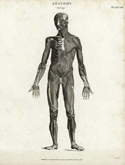
Background imageAnatomist Collection: Anatomy of human musculature showing outer muscles from the front, 1820 (copperplate engraving)

Anatomy of human musculature showing outer muscles from the front, 1820 (copperplate engraving)
FLO4591432 Anatomy of human musculature showing outer muscles from the front, 1820 (copperplate engraving); (add.info.: Anatomy of human musculature showing outer muscles from the front)

Background imageAnatomist Collection: Anatomy of human musculature from the back showing outer muscles, 1820 (copperplate engraving)

Anatomy of human musculature from the back showing outer muscles, 1820 (copperplate engraving)
FLO4591171 Anatomy of human musculature from the back showing outer muscles, 1820 (copperplate engraving); (add.info.: Anatomy of human musculature from the back showing outer muscles)

Background imageAnatomist Collection: Anatomy of human internal organs from the front showing heart, stomach, intestines

Anatomy of human internal organs from the front showing heart, stomach, intestines, kidneys and bladder
FLO4590679 Anatomy of human internal organs from the front showing heart, stomach, intestines, kidneys and bladder. Copperplate engraving by Milton from Abraham Rees' Cyclopedia or Universal

Background imageAnatomist Collection: Head muscles. Lithograph of Lebrun, based on a drawing by Leveille

Head muscles. Lithograph of Lebrun, based on a drawing by Leveille, in Petity Atlas complet d'Anatomie descriptive
FLO4672607 Head muscles. Lithograph of Lebrun, based on a drawing by Leveille, in Petity Atlas complet d'Anatomie descriptive du Corps Humain, by Dr

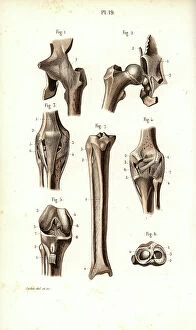
Background imageAnatomist Collection: Hip joint and thigh bone. Lithograph of Corbie, in Petity Atlas complet d'Anatomie descriptive du

Hip joint and thigh bone. Lithograph of Corbie, in Petity Atlas complet d'Anatomie descriptive du Corps Humain
FLO4672595 Hip joint and thigh bone. Lithograph of Corbie, in Petity Atlas complet d'Anatomie descriptive du Corps Humain, by Dr

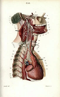
Background imageAnatomist Collection: Sympathic nervous system in the thoracic, lumbar and sacral plexus. Lithograph by Davesne

Sympathic nervous system in the thoracic, lumbar and sacral plexus. Lithograph by Davesne
FLO4672529 Sympathic nervous system in the thoracic, lumbar and sacral plexus. Lithograph by Davesne, based on a drawing by Leveille

Background imageAnatomist Collection: Sympathic nervous system in the cardiac plexus. Lithograph by Davesne

Sympathic nervous system in the cardiac plexus. Lithograph by Davesne, based on a drawing by Leveille
FLO4672521 Sympathic nervous system in the cardiac plexus. Lithograph by Davesne, based on a drawing by Leveille, in Petiure Atlas complet d'Anatomie descriptive du Corps Humain, by Dr

Background imageAnatomist Collection: Nervous system of the back. Lithograph of Corbie, based on a drawing by Leveille

Nervous system of the back. Lithograph of Corbie, based on a drawing by Leveille
FLO4672457 Nervous system of the back. Lithograph of Corbie, based on a drawing by Leveille, in Petity Atlas complet d'Anatomie descriptive du Corps Humain, by Dr

Background imageAnatomist Collection: Facial and auditory nerves. Lithograph by Davesne, based on a drawing by Leveille

Facial and auditory nerves. Lithograph by Davesne, based on a drawing by Leveille
FLO4672442 Facial and auditory nerves. Lithograph by Davesne, based on a drawing by Leveille, in Petite Atlas complet d'Anatomie descriptive du Corps Humain, by Dr

Background imageAnatomist Collection: Intercostal nerves. Lithograph of Corbie, based on a drawing by Leveille

Intercostal nerves. Lithograph of Corbie, based on a drawing by Leveille, in Petity Atlas complet d'Anatomie
FLO4672415 Intercostal nerves. Lithograph of Corbie, based on a drawing by Leveille, in Petity Atlas complet d'Anatomie descriptive du Corps Humain, by Dr

Background imageAnatomist Collection: Omnium Animalum : Omnium Animalum, mixed strange animal kingdom creatures

Omnium Animalum : Omnium Animalum, mixed strange animal kingdom creatures
UIG5327222 Omnium Animalum : Omnium Animalum, mixed strange animal kingdom creatures. From the 1642 book Monstrorum Historia by Ulisse Aldrovandi (Bologna, 1522-1605)

Background imageAnatomist Collection: Animal Tigridi Affine : Animal Tigridi affine: an animal akin to a tiger

Animal Tigridi Affine : Animal Tigridi affine: an animal akin to a tiger. From the 1642 book Monstrorum Historia by
UIG5327220 Animal Tigridi Affine : Animal Tigridi affine: an animal akin to a tiger. From the 1642 book Monstrorum Historia by Ulisse Aldrovandi (Bologna, 1522-1605)

Background imageAnatomist Collection: Chicken Man : Human and chicken combined creatures. From the 1642 book Monstrorum Historia by

Chicken Man : Human and chicken combined creatures. From the 1642 book Monstrorum Historia by Ulisse Aldrovandi
UIG5327206 Chicken Man : Human and chicken combined creatures. From the 1642 book Monstrorum Historia by Ulisse Aldrovandi (Bologna, 1522-1605)

Background imageAnatomist Collection: Herinaceus Marinus : Herinaceus Marinus. From the 1642 book Monstrorum Historia by Ulisse

Herinaceus Marinus : Herinaceus Marinus. From the 1642 book Monstrorum Historia by Ulisse Aldrovandi (Bologna)
UIG5327205 Herinaceus Marinus : Herinaceus Marinus. From the 1642 book Monstrorum Historia by Ulisse Aldrovandi (Bologna, 1522-1605)

Background imageAnatomist Collection: Rana piscatrix ala feu Bufo Marinus : Rana piscatrix ala feu Bufo Marinus

Rana piscatrix ala feu Bufo Marinus : Rana piscatrix ala feu Bufo Marinus
UIG5327202 Rana piscatrix ala feu Bufo Marinus : Rana piscatrix ala feu Bufo Marinus. Rana piscatrix is a type of anglerfish or monkfish today called Lophius piscatorius

Background imageAnatomist Collection: Andora Piscis aspectu fuillo : Andora Piscis aspectu fuillo. A sea cow like hairy fish monster

Andora Piscis aspectu fuillo : Andora Piscis aspectu fuillo. A sea cow like hairy fish monster
UIG5327201 Andora Piscis aspectu fuillo : Andora Piscis aspectu fuillo. A sea cow like hairy fish monster, from the 1642 book Monstrorum Historia by Ulisse Aldrovandi (Bologna, 1522-1605)

Background imageAnatomist Collection: Seal & Walrus Creatures : Seal & Walrus Creatures. From the 1642 book Monstrorum Historia by

Seal & Walrus Creatures : Seal & Walrus Creatures. From the 1642 book Monstrorum Historia by Ulisse Aldrovandi
UIG5327193 Seal & Walrus Creatures : Seal & Walrus Creatures. From the 1642 book Monstrorum Historia by Ulisse Aldrovandi (Bologna, 1522-1605)

Background imageAnatomist Collection: Human Monsters : Six people with deformities. From the 1642 book Monstrorum Historia by Ulisse

Human Monsters : Six people with deformities. From the 1642 book Monstrorum Historia by Ulisse Aldrovandi (Bologna)
UIG5327179 Human Monsters : Six people with deformities. From the 1642 book Monstrorum Historia by Ulisse Aldrovandi (Bologna, 1522-1605)

Background imageAnatomist Collection: Monstrum biceps & septiceps : Monstrum biceps & septiceps. Multi armed men

Monstrum biceps & septiceps : Monstrum biceps & septiceps. Multi armed men
UIG5327177 Monstrum biceps & septiceps : Monstrum biceps & septiceps. Multi armed men. From the 1642 book Monstrorum Historia by Ulisse Aldrovandi (Bologna, 1522-1605)

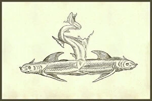
Background imageAnatomist Collection: Piscis Dicephalos : Piscis Dicephalos, two headed fish. From the 1642 book Monstrorum Historia by

Piscis Dicephalos : Piscis Dicephalos, two headed fish. From the 1642 book Monstrorum Historia by Ulisse Aldrovandi
UIG5327152 Piscis Dicephalos : Piscis Dicephalos, two headed fish. From the 1642 book Monstrorum Historia by Ulisse Aldrovandi (Bologna, 1522-1605)

Background imageAnatomist Collection: Capra Humana Facie : Capra Humana Facie. From the 1642 book Monstrorum Historia by Ulisse

Capra Humana Facie : Capra Humana Facie. From the 1642 book Monstrorum Historia by Ulisse Aldrovandi (Bologna)
UIG5327147 Capra Humana Facie : Capra Humana Facie. From the 1642 book Monstrorum Historia by Ulisse Aldrovandi (Bologna, 1522-1605)

Background imageAnatomist Collection: Puer capite elephantino : Puer capite elephantino, boy with an elephant's head, elephant man

Puer capite elephantino : Puer capite elephantino, boy with an elephant's head, elephant man
UIG5327138 Puer capite elephantino : Puer capite elephantino, boy with an elephant's head, elephant man. From the 1642 book Monstrorum Historia by Ulisse Aldrovandi (Bologna, 1522-1605)

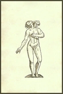
Background imageAnatomist Collection: Two heads on one body, 1642 (engraving)

Two heads on one body, 1642 (engraving)
UIG5327120 Two heads on one body, 1642 (engraving); (add.info.: Monstrum hermaphroditicum from the 1642 book "Monstrorum Historia" by Ulisse Aldrovandi (Bologna)

Background imageAnatomist Collection: Infans biceps obscaeno membro : Infans biceps obscaeno membro, 407, two heads on one body

Infans biceps obscaeno membro : Infans biceps obscaeno membro, 407, two heads on one body, siamese twins
UIG5327115 Infans biceps obscaeno membro : Infans biceps obscaeno membro, 407, two heads on one body, siamese twins. From the 1642 book Monstrorum Historia by Ulisse Aldrovandi (Bologna, 1522-1605)

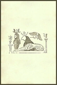
Background imageAnatomist Collection: Gryphis Tertia (Griffin) : The griffin, griffon, or gryphon is a legendary creature with the body

Gryphis Tertia (Griffin) : The griffin, griffon, or gryphon is a legendary creature with the body, tail
UIG5327074 Gryphis Tertia (Griffin) : The griffin, griffon, or gryphon is a legendary creature with the body, tail, and back legs of a lion; the head and wings of an eagle;


All Professionally Made to Order for Quick Shipping

Anatomists have played a crucial role in unraveling the mysteries of the human body throughout history. One such pioneer was Robert Burton, whose extensive research and writings on anatomy left an indelible mark on the field. Another notable figure was EUSTACHIO Bartolomeo, an Italian anatomist who made significant contributions during the 16th century. Intriguingly, a lithograph titled "Odin questioning Mimir" captures the essence of curiosity that drives anatomists to seek knowledge. This thirst for understanding is exemplified by Sir Richard Owen, a renowned British anatomist from the 19th century whose work continues to inspire modern scientists. Richard Owen's dedication to his craft is evident in various depictions of him throughout history. In one caricature from Vanity Fair in 1873, aptly titled "Old bones, " he is portrayed as a distinguished scholar with wisdom etched onto his face. An image from his study at BMNH in 1883 showcases his meticulous approach towards dissecting and studying specimens. The intricate details captured in engravings like "The Sacred Heart of Jesus" or "The pollen of plants" demonstrate how anatomists explore not only human anatomy but also delve into other aspects of nature's design. Richard Owen himself had a profound impact on paleontology, as depicted in C016 / 5008 - an image that pays homage to his groundbreaking discoveries. Throughout history, heart anatomists have held a special place within this scientific realm. Figures like Guillaume Dupuytren and William Hunter dedicated their lives to unraveling the complexities of this vital organ. Their contributions paved the way for advancements in cardiac medicine that continue to save lives today. From ancient times until now, these brilliant minds have shaped our understanding of ourselves and our world through their tireless pursuit of knowledge about our bodies' inner workings. The legacy they leave behind serves as an inspiration for future generations aspiring to unravel the secrets of human anatomy.