Fraud Blocker Skip to main content

Anatomist Collection (page 7)

Anatomists have played a crucial role in unraveling the mysteries of the human body throughout history

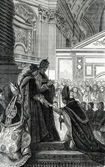
Background imageAnatomist Collection: The anatomist Niels Stensen (Stenon or Steensen or Steno or stenone) (1638-1688)

The anatomist Niels Stensen (Stenon or Steensen or Steno or stenone) (1638-1688)
ELD4905480 The anatomist Niels Stensen (Stenon or Steensen or Steno or stenone) (1638-1688) crowned of Titiopolis on 19/09/1677 by Pope Innocent XI and Cardinal Barbarigo

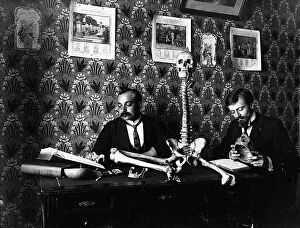
Background imageAnatomist Collection: France, Pays de la Loire, Maine-et-Loire (49), Angers: hospital of Angers, medical school

France, Pays de la Loire, Maine-et-Loire (49), Angers: hospital of Angers, medical school
LUX4771257 France, Pays de la Loire, Maine-et-Loire (49), Angers: hospital of Angers, medical school, medical students study anatomy with the help of a skeleton and the book

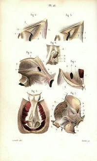
Background imageAnatomist Collection: External female parts of generation, labia, clitoris, perineum, and anus

External female parts of generation, labia, clitoris, perineum, and anus. Copperplate engraving by Andrew Bell after an
FLO4696243 External female parts of generation, labia, clitoris, perineum, and anus. Copperplate engraving by Andrew Bell after an illustration by Jan van Rymsdyk from William Smellie's A Set of

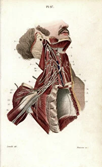
Background imageAnatomist Collection: Deep nervous system of the arm. Lithograph by Davesne, based on a drawing by Leveille

Deep nervous system of the arm. Lithograph by Davesne, based on a drawing by Leveille
FLO4673176 Deep nervous system of the arm. Lithograph by Davesne, based on a drawing by Leveille, in Petity Atlas complet d'Anatomie descriptive du Corps Humain, by Dr

Background imageAnatomist Collection: Brain, pons, spinal bulb, pineal gland etc... Lithograph by Davesne, based on a drawing by Leveille

Brain, pons, spinal bulb, pineal gland etc... Lithograph by Davesne, based on a drawing by Leveille
FLO4673157 Brain, pons, spinal bulb, pineal gland etc... Lithograph by Davesne, based on a drawing by Leveille, in Petity Atlas complet d'Anatomie descriptive du Corps Humain, by Dr

Background imageAnatomist Collection: Brachial plexus. Lithograph by Davesne, based on a drawing by Leveille

Brachial plexus. Lithograph by Davesne, based on a drawing by Leveille, in Petity Atlas complet d'Anatomie
FLO4673154 Brachial plexus. Lithograph by Davesne, based on a drawing by Leveille, in Petity Atlas complet d'Anatomie descriptive du Corps Humain, by Dr

Background imageAnatomist Collection: Section through the brain. Lithograph by Davesne, based on a drawing by Leveille

Section through the brain. Lithograph by Davesne, based on a drawing by Leveille
FLO4673112 Section through the brain. Lithograph by Davesne, based on a drawing by Leveille, in Petity Atlas complet d'Anatomie descriptive du Corps Humain, by Dr

Background imageAnatomist Collection: Different sections of the skull. Lithograph of Corbie, based on a drawing by Leveille

Different sections of the skull. Lithograph of Corbie, based on a drawing by Leveille
FLO4673087 Different sections of the skull. Lithograph of Corbie, based on a drawing by Leveille, in Petity Atlas complet d'Anatomie descriptive du Corps Humain, by Dr

Background imageAnatomist Collection: Lymph nodes and vessels of the head, neck and chest. Lithograph of Corbie

Lymph nodes and vessels of the head, neck and chest. Lithograph of Corbie, based on a drawing by Leveille
FLO4673058 Lymph nodes and vessels of the head, neck and chest. Lithograph of Corbie, based on a drawing by Leveille, in Petity Atlas complet d'Anatomie descriptive du Corps Humain, by Dr

Background imageAnatomist Collection: Lymph nodes and deep vessels of the leg. Lithograph of Corbie, based on a drawing by Leveille

Lymph nodes and deep vessels of the leg. Lithograph of Corbie, based on a drawing by Leveille
FLO4673028 Lymph nodes and deep vessels of the leg. Lithograph of Corbie, based on a drawing by Leveille, in Petity Atlas complet d'Anatomie descriptive du Corps Humain, by Dr

Background imageAnatomist Collection: Sinus or skull cavites. Humerus and vertebrae. Lithograph of Corbie, based on a drawing by Leveille

Sinus or skull cavites. Humerus and vertebrae. Lithograph of Corbie, based on a drawing by Leveille
FLO4673010 Sinus or skull cavites. Humerus and vertebrae. Lithograph of Corbie, based on a drawing by Leveille, in Petity Atlas complet d'Anatomie descriptive du Corps Humain, by Dr

Background imageAnatomist Collection: Arm and hand circulatory system. Lithograph of Corbie, based on a drawing by Leveille

Arm and hand circulatory system. Lithograph of Corbie, based on a drawing by Leveille
FLO4672987 Arm and hand circulatory system. Lithograph of Corbie, based on a drawing by Leveille, in Petity Atlas complet d'Anatomie descriptive du Corps Humain, by Dr

Background imageAnatomist Collection: Circulatory system of the leg and foot. Lithograph of Choubard, based on a drawing by Leveille

Circulatory system of the leg and foot. Lithograph of Choubard, based on a drawing by Leveille
FLO4672981 Circulatory system of the leg and foot. Lithograph of Choubard, based on a drawing by Leveille, in Petity Atlas complet d'Anatomie descriptive du Corps Humain, by Dr

Background imageAnatomist Collection: Circulatory system of the leg and hand. Lithograph by Davesne, based on a drawing by Leveille

Circulatory system of the leg and hand. Lithograph by Davesne, based on a drawing by Leveille
FLO4672964 Circulatory system of the leg and hand. Lithograph by Davesne, based on a drawing by Leveille, in Petity Atlas complet d'Anatomie descriptive du Corps Humain, by Dr

Background imageAnatomist Collection: The circulatory system of the stomach, liver, kidneys and pancreas. Lithograph of Choubard

The circulatory system of the stomach, liver, kidneys and pancreas. Lithograph of Choubard
FLO4672920 The circulatory system of the stomach, liver, kidneys and pancreas. Lithograph of Choubard, based on a drawing by Leveille

Background imageAnatomist Collection: Jaw bone. Lithograph of Corbie, in Petity Atlas complet d'Anatomie descriptive du Corps Humain

Jaw bone. Lithograph of Corbie, in Petity Atlas complet d'Anatomie descriptive du Corps Humain, by Dr
FLO4672890 Jaw bone. Lithograph of Corbie, in Petity Atlas complet d'Anatomie descriptive du Corps Humain, by Dr. Joseph Nicolas Masse, published by Mequignon Marvis, Paris (France)

Background imageAnatomist Collection: Spleen, kidneys, caecum, bile and pancreas. Lithograph of Corbie, based on a drawing by Leveille

Spleen, kidneys, caecum, bile and pancreas. Lithograph of Corbie, based on a drawing by Leveille
FLO4672861 Spleen, kidneys, caecum, bile and pancreas. Lithograph of Corbie, based on a drawing by Leveille, in Petiure Atlas complet d'Anatomie descriptive du Corps Humain, by Dr

Background imageAnatomist Collection: Ear and eardrum. Lithograph of Corbie, based on a drawing by Leveille

Ear and eardrum. Lithograph of Corbie, based on a drawing by Leveille, in Petity Atlas complet d'Anatomie
FLO4672822 Ear and eardrum. Lithograph of Corbie, based on a drawing by Leveille, in Petity Atlas complet d'Anatomie descriptive du Corps Humain, by Dr

Background imageAnatomist Collection: Sections of the nasal cavity. Lithograph of Corbie, based on a drawing by Leveille
Sections of the nasal cavity. Lithograph of Corbie, based on a drawing by Leveille
FLO4672810 Sections of the nasal cavity. Lithograph of Corbie, based on a drawing by Leveille, in Petity Atlas complet d'Anatomie descriptive du Corps Humain, by Dr

Background imageAnatomist Collection: Aponevrosis of the abdomen and perinee. Lithograph of Corbie, based on a drawing by Leveille

Aponevrosis of the abdomen and perinee. Lithograph of Corbie, based on a drawing by Leveille
FLO4672789 Aponevrosis of the abdomen and perinee. Lithograph of Corbie, based on a drawing by Leveille, in Petity Atlas complet d'Anatomie descriptive du Corps Humain, by Dr

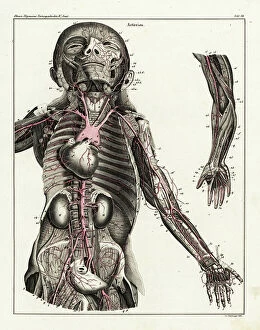
Background imageAnatomist Collection: Anatomy of the human arterial system in the upper torso. Handcoloured lithograph by St

Anatomy of the human arterial system in the upper torso. Handcoloured lithograph by St
FLO4684926 Anatomy of the human arterial system in the upper torso. Handcoloured lithograph by St. Schillinger from Lorenz Oken's Universal Natural History

Background imageAnatomist Collection: Anatomy of human venous return system of venal valves in the upper torso

Anatomy of human venous return system of venal valves in the upper torso. Lithograph from Lorenz Oken's Universal
FLO4684841 Anatomy of human venous return system of venal valves in the upper torso. Lithograph from Lorenz Oken's Universal Natural History, Allgemeine Naturgeschichte fur alle Stande

Background imageAnatomist Collection: Jules II (1443-1513) - engraving of 1823 in ' Drolatiques Songes de Pantagruel' by Francois Rabelais

Jules II (1443-1513) - engraving of 1823 in " Drolatiques Songes de Pantagruel" by Francois Rabelais
JEB4934033 Jules II (1443-1513) - engraving of 1823 in " Drolatiques Songes de Pantagruel" by Francois Rabelais (1494-1553) - (The Capricious Dreams of Pantagruel)

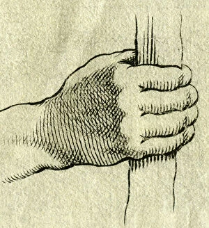
Background imageAnatomist Collection: a packing hand, copperplate engraving, about 1700

a packing hand, copperplate engraving, about 1700
UTD4922026 a packing hand, copperplate engraving, about 1700; (add.info.: a packing hand, copperplate engraving, about 1700 ©HistAuge/United Archives/Leemage); United Archives/HistAuge.

Background imageAnatomist Collection: head with curly hair, copperplate engraving , about 1700

head with curly hair, copperplate engraving , about 1700
UTD4921906 head with curly hair, copperplate engraving , about 1700; (add.info.: head with curly hair, copperplate engraving)

Background imageAnatomist Collection: Head of a viper with bark, c.1800-1850 (object)

Head of a viper with bark, c.1800-1850 (object)
7418311 Head of a viper with bark, c.1800-1850 (object); Ecole Nationale Veterinaire, Toulouse, France; (add.info.: Bark viper head: demountable model of clastic anatomy made of papier-mache, cork)

Background imageAnatomist Collection: Engraving depicting the bones of the human foot, 19th century

Engraving depicting the bones of the human foot, 19th century
5309455 Engraving depicting the bones of the human foot, 19th century; (add.info.: Engraving depicting the bones of the human foot. Dated 19th century 01/02/1850); Universal History Archive/UIG.

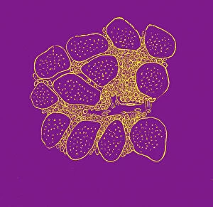
Background imageAnatomist Collection: Cross section of a a muscle with cancer, 1867 (engraving)

Cross section of a a muscle with cancer, 1867 (engraving)
8766935 Cross section of a a muscle with cancer, 1867 (engraving); Private Collection; (add.info.: Cross section of a a muscle with cancer. ( Dr. Jaccoud dictionary . 1867)

Background imageAnatomist Collection: Thomas Henry Huxley, 1825 - 1895

Thomas Henry Huxley, 1825 - 1895
3083225 Thomas Henry Huxley, 1825 - 1895.; Private Collection; (add.info.: English biologist aka "Darwin's Bulldog" for his advocacy of Charles Darwin's theory of evolution)

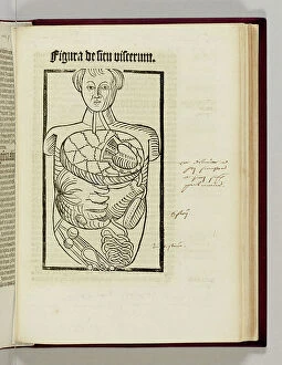
Background imageAnatomist Collection: A page from 'Antropologium de Hominis Dignitate, Natura, et Proprietatibus ee Elementis' by Magnus

A page from 'Antropologium de Hominis Dignitate, Natura, et Proprietatibus ee Elementis' by Magnus Hundt
1764829 A page from 'Antropologium de Hominis Dignitate, Natura, et Proprietatibus ee Elementis' by Magnus Hundt, the Elder (1449-1519), 1501 (woodcut print) by Hundt

Background imageAnatomist Collection: Albrecht von Haller, 1874 (engraving)

Albrecht von Haller, 1874 (engraving)
1574758 Albrecht von Haller, 1874 (engraving); (add.info.: Albrecht von Haller (1708-1777) Swiss anatomist and physiologist. Engraving, Paris, 1874.); Universal History Archive/UIG.

Background imageAnatomist Collection: William Sharpey, 1869

William Sharpey, 1869
1577008 William Sharpey, 1869; (add.info.: William Sharpey (1 April 1802 - 11 April 1880) was a Scottish anatomist and physiologist

Background imageAnatomist Collection: Proportions of the human body after Leonardo's studies, also called Vitruvian man

Proportions of the human body after Leonardo's studies, also called Vitruvian man
528748 Proportions of the human body after Leonardo's studies, also called Vitruvian man. Male human body with limbs extended, superimposed on a grid and a circle

Background imageAnatomist Collection: How man learns from the human eye (litho)

How man learns from the human eye (litho)
8676259 How man learns from the human eye (litho) by English School, (20th century); Private Collection; (add.info.: How man learns from the human eye. Inventions using lenses)

Background imageAnatomist Collection: Oxford: Anatomical School (engraving)

Oxford: Anatomical School (engraving)
8675443 Oxford: Anatomical School (engraving) by English School, (19th century); Private Collection; (add.info.: Anatomical School)

Background imageAnatomist Collection: Votaries of the Weed No 3 (colour litho)

Votaries of the Weed No 3 (colour litho)
8660493 Votaries of the Weed No 3 (colour litho) by Clarke, Joseph Clayton (Kyd) (1856-1937); Private Collection; (add.info.: Votaries of the Weed, No 3)

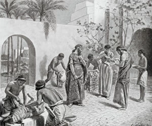
Background imageAnatomist Collection: Hepatoscopy, or Divination by the Liver (litho)

Hepatoscopy, or Divination by the Liver (litho)
8660936 Hepatoscopy, or Divination by the Liver (litho) by Weatherstone, A.C. (fl.1888-1929); Private Collection; (add.info.: Hepatoscopy, or Divination by the Liver)

Background imageAnatomist Collection: Development of muscles through exercise (coloured engraving)

Development of muscles through exercise (coloured engraving)
8655710 Development of muscles through exercise (coloured engraving) by French School, (19th century) (after); Private Collection; (add.info.: Development of muscles through exercise)

Background imageAnatomist Collection: Normal position of twins in the womb during pregnancy (coloured engraving)

Normal position of twins in the womb during pregnancy (coloured engraving)
8655687 Normal position of twins in the womb during pregnancy (coloured engraving) by French School, (19th century) (after); Private Collection; (add.info)

Background imageAnatomist Collection: Interior of the nose (coloured engraving)

Interior of the nose (coloured engraving)
8655758 Interior of the nose (coloured engraving) by French School, (19th century) (after); Private Collection; (add.info.: Interior of the nose)

Background imageAnatomist Collection: Organs of the digestive system (coloured engraving)

Organs of the digestive system (coloured engraving)
8655750 Organs of the digestive system (coloured engraving) by French School, (19th century) (after); Private Collection; (add.info.: Organs of the digestive system)

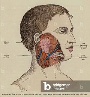
Background imageAnatomist Collection: Saivary glands and muscles, nerves and blood vessels of the cheek (coloured engraving)

Saivary glands and muscles, nerves and blood vessels of the cheek (coloured engraving)
8655749 Saivary glands and muscles, nerves and blood vessels of the cheek (coloured engraving) by French School, (19th century) (after); Private Collection; (add.info.: Saivary glands and muscles)

Background imageAnatomist Collection: Section of the small intestine showing the structure of the mucosa (coloured engraving)

Section of the small intestine showing the structure of the mucosa (coloured engraving)
8655747 Section of the small intestine showing the structure of the mucosa (coloured engraving) by French School, (19th century) (after); Private Collection; (add.info)

Background imageAnatomist Collection: Blood vessels of the neck (coloured engraving)

Blood vessels of the neck (coloured engraving)
8655731 Blood vessels of the neck (coloured engraving) by French School, (19th century) (after); Private Collection; (add.info.: Blood vessels of the neck)

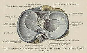
Background imageAnatomist Collection: Upper End of Tibia, with Menisci and Attached Portions of Crucial Ligaments (colour litho)

Upper End of Tibia, with Menisci and Attached Portions of Crucial Ligaments (colour litho)
8667637 Upper End of Tibia, with Menisci and Attached Portions of Crucial Ligaments (colour litho) by English School, (20th century); Private Collection; (add.info.: Upper End of Tibia)

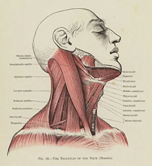
Background imageAnatomist Collection: The Triangles of the Neck, Muscles (colour litho)

The Triangles of the Neck, Muscles (colour litho)
8667618 The Triangles of the Neck, Muscles (colour litho) by English School, (20th century); Private Collection; (add.info.: The Triangles of the Neck, Muscles)

Background imageAnatomist Collection: Superficial Muscles on the Front of the Arm and Forearm (colour litho)

Superficial Muscles on the Front of the Arm and Forearm (colour litho)
8667617 Superficial Muscles on the Front of the Arm and Forearm (colour litho) by English School, (20th century); Private Collection; (add.info)

Background imageAnatomist Collection: The Front of the Body (colour litho)

The Front of the Body (colour litho)
8667613 The Front of the Body (colour litho) by English School, (20th century); Private Collection; (add.info.: The Front of the Body)


All Professionally Made to Order for Quick Shipping

Anatomists have played a crucial role in unraveling the mysteries of the human body throughout history. One such pioneer was Robert Burton, whose extensive research and writings on anatomy left an indelible mark on the field. Another notable figure was EUSTACHIO Bartolomeo, an Italian anatomist who made significant contributions during the 16th century. Intriguingly, a lithograph titled "Odin questioning Mimir" captures the essence of curiosity that drives anatomists to seek knowledge. This thirst for understanding is exemplified by Sir Richard Owen, a renowned British anatomist from the 19th century whose work continues to inspire modern scientists. Richard Owen's dedication to his craft is evident in various depictions of him throughout history. In one caricature from Vanity Fair in 1873, aptly titled "Old bones, " he is portrayed as a distinguished scholar with wisdom etched onto his face. An image from his study at BMNH in 1883 showcases his meticulous approach towards dissecting and studying specimens. The intricate details captured in engravings like "The Sacred Heart of Jesus" or "The pollen of plants" demonstrate how anatomists explore not only human anatomy but also delve into other aspects of nature's design. Richard Owen himself had a profound impact on paleontology, as depicted in C016 / 5008 - an image that pays homage to his groundbreaking discoveries. Throughout history, heart anatomists have held a special place within this scientific realm. Figures like Guillaume Dupuytren and William Hunter dedicated their lives to unraveling the complexities of this vital organ. Their contributions paved the way for advancements in cardiac medicine that continue to save lives today. From ancient times until now, these brilliant minds have shaped our understanding of ourselves and our world through their tireless pursuit of knowledge about our bodies' inner workings. The legacy they leave behind serves as an inspiration for future generations aspiring to unravel the secrets of human anatomy.