Fraud Blocker Skip to main content

Anatomist Collection (page 9)

Anatomists have played a crucial role in unraveling the mysteries of the human body throughout history

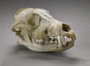
Background imageAnatomist Collection: Canis lupus (Wolf gris - gray (grey) woolf or timber woolf), Museum d'histoire naturelle de

Canis lupus (Wolf gris - gray (grey) woolf or timber woolf), Museum d'histoire naturelle de Marseille
CSO5006426 Canis lupus (Wolf gris - gray (grey) woolf or timber woolf), Museum d'histoire naturelle de Marseille; (add.info.: Canis lupus (Wolf gris - gray (grey) woolf or timber woolf)

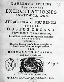
Background imageAnatomist Collection: Frontispice of ' Exercisitationes anatomicae duae de structura et sus renum' of 1725 by Lorenzo

Frontispice of " Exercisitationes anatomicae duae de structura et sus renum" of 1725 by Lorenzo (Laurentio)
XEE5028108 Frontispice of " Exercisitationes anatomicae duae de structura et sus renum" of 1725 by Lorenzo (Laurentio) Bellini (Firenze 1643 - Pisa 1704) - Biblioteca nazionale braidense

Background imageAnatomist Collection: Portrait of Bernard Siegfried Albinus (1697-1770) German anatomist

Portrait of Bernard Siegfried Albinus (1697-1770) German anatomist
XEE5027659 Portrait of Bernard Siegfried Albinus (1697-1770) German anatomist; (add.info.: Portrait of Bernard Siegfried Albinus (1697-1770) German anatomist); Stefano Bianchetti.

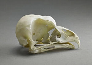
Background imageAnatomist Collection: Bubo bubo (Skull of the Eurasian eagle-owl), Museum of Natural History of Marseille

Bubo bubo (Skull of the Eurasian eagle-owl), Museum of Natural History of Marseille
CSO5006276 Bubo bubo (Skull of the Eurasian eagle-owl), Museum of Natural History of Marseille; (add.info.: Bubo bubo (Skull of the Eurasian eagle-owl)

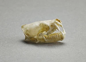
Background imageAnatomist Collection: Vesperugo serotinus (Chiroptere) (Crane de la grande serotine ou serotine commune - skull of an)

Vesperugo serotinus (Chiroptere) (Crane de la grande serotine ou serotine commune - skull of an european long-eared bat)
CSO5006273 Vesperugo serotinus (Chiroptere) (Crane de la grande serotine ou serotine commune - skull of an european long-eared bat or European brown bat)

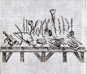
Background imageAnatomist Collection: Instrument surgical csurgical instruments) - in ' De anatomome corporis humani' by Guido Guidi

Instrument surgical csurgical instruments) - in " De anatomome corporis humani" by Guido Guidi (Vidus Vidius)
XEE5029681 Instrument surgical csurgical instruments) - in " De anatomome corporis humani" by Guido Guidi (Vidus Vidius) (1509-1569) Italian surgeon and anatomist

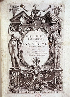
Background imageAnatomist Collection: Frontispice of ' De anatomome corporis humani' by Guido Guidi (Vidus Vidius) (1509-1569)

Frontispice of " De anatomome corporis humani" by Guido Guidi (Vidus Vidius) (1509-1569)
XEE5029679 Frontispice of " De anatomome corporis humani" by Guido Guidi (Vidus Vidius) (1509-1569) Italian surgeon and anatomist

Background imageAnatomist Collection: Anatomy: representation of the uterus by Garioponto (970 - 1050) Italian doctor of the Middle Ages

Anatomy: representation of the uterus by Garioponto (970 - 1050) Italian doctor of the Middle Ages
XEE5029571 Anatomy: representation of the uterus by Garioponto (970 - 1050) Italian doctor of the Middle Ages, Biblioteca Civica Bertoliana

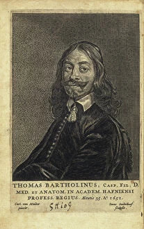
Background imageAnatomist Collection: BARTHOLIN, Thomas (1616-1680). Danish physician, mathematician, and theologian

BARTHOLIN, Thomas (1616-1680). Danish physician, mathematician, and theologian
CUL4847818 BARTHOLIN, Thomas (1616-1680). Danish physician, mathematician, and theologian. He is best known for his work in the discovery of the lymphatic system in humans

Background imageAnatomist Collection: Front view of a healthy female pelvis. Copperplate engraving by Andrew Bell after an illustration

Front view of a healthy female pelvis. Copperplate engraving by Andrew Bell after an illustration by Jan van Rymsdyk
FLO4695855 Front view of a healthy female pelvis. Copperplate engraving by Andrew Bell after an illustration by Jan van Rymsdyk from William Smellie's A Set of Anatomical Tables, Charles Elliot

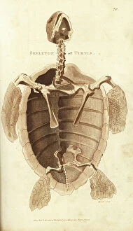
Background imageAnatomist Collection: Skeleton of a turtle. Handcoloured copperplate engraving by Heath after an illustration by George

Skeleton of a turtle. Handcoloured copperplate engraving by Heath after an illustration by George Shaw from his General
FLO4694475 Skeleton of a turtle. Handcoloured copperplate engraving by Heath after an illustration by George Shaw from his General Zoology, Amphibia, London, 1801.; (add.info.: Skeleton of a turtle)

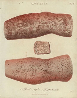
Background imageAnatomist Collection: La scarlatine ou feevre ecarlate - Lithograph by John Pass, published in 'Encyclopedia

La scarlatine ou feevre ecarlate - Lithograph by John Pass, published in "Encyclopedia Londinensis" by John
FLO4618755 La scarlatine ou feevre ecarlate - Lithograph by John Pass, published in "Encyclopedia Londinensis" by John Wilkes, edition J.Adlard, London

Background imageAnatomist Collection: Skin Diseases: Nodular or Hyde Prurigo and Varietes of Psoriasis - Lithography by John Pass

Skin Diseases: Nodular or Hyde Prurigo and Varietes of Psoriasis - Lithography by John Pass
FLO4618824 Skin Diseases: Nodular or Hyde Prurigo and Varietes of Psoriasis - Lithography by John Pass, published in "Encyclopedia Londinensis" by John Wilkes, J.Adlard Edition, London

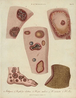
Background imageAnatomist Collection: Ichthyosis (peeling), dyshidrosis (bullous eczema) and herpetic skin diseases -Lithography by John

Ichthyosis (peeling), dyshidrosis (bullous eczema) and herpetic skin diseases -Lithography by John Pass
FLO4618812 Ichthyosis (peeling), dyshidrosis (bullous eczema) and herpetic skin diseases -Lithography by John Pass, published in "Encyclopedia Londinensis" by John Wilkes, J.Adlard edition

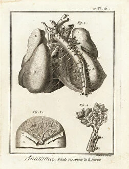
Background imageAnatomist Collection: Arteries in the chest cavity to the heart and lungs. Copperplate engraving by Robert Benard after

Arteries in the chest cavity to the heart and lungs. Copperplate engraving by Robert Benard after an illustration by
FLO4611670 Arteries in the chest cavity to the heart and lungs. Copperplate engraving by Robert Benard after an illustration by Albrecht von Haller from Denis Diderot's Encyclopedia, Pellet

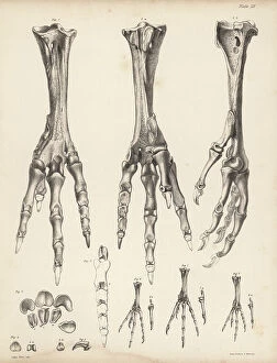
Background imageAnatomist Collection: Metatarsus and toes of the dodo, Raphus cucullatus 1-5, foot of the yellow-footed green pigeon

Metatarsus and toes of the dodo, Raphus cucullatus 1-5, foot of the yellow-footed green pigeon, Treron chlorigaster 6
FLO4612178 Metatarsus and toes of the dodo, Raphus cucullatus 1-5, foot of the yellow-footed green pigeon, Treron chlorigaster 6, stock dove, Columba oenas 7 and partridge pigeon

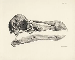
Background imageAnatomist Collection: Side view of the skull of a dodo. Lithograph from Hugh Edwin Strickland

Side view of the skull of a dodo. Lithograph from Hugh Edwin Strickland and Alexander Gordon Melville's The Dodo
FLO4612152 Side view of the skull of a dodo. Lithograph from Hugh Edwin Strickland and Alexander Gordon Melville's The Dodo and its Kindred, London, Reeve, Benham and Reeve

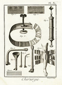
Background imageAnatomist Collection: Sutures, withers, fine-tip syringe by Dominique Anel, siphon, injection tubes

Sutures, withers, fine-tip syringe by Dominique Anel, siphon, injection tubes
FLO4611953 Sutures, withers, fine-tip syringe by Dominique Anel, siphon, injection tubes, 18-sheet bandage for complex fractures

Background imageAnatomist Collection: France: Medical photograph of a spinal malformation, 1890

France: Medical photograph of a spinal malformation, 1890
LUX4755402 France: Medical photograph of a spinal malformation, 1890; (add.info.: France: Medical photograph of a spinal malformation, 1890); Photo © Lux-in-Fine.

Background imageAnatomist Collection: Bertillonage (Bertillon system or forensic anthropometry) a forensic technique developed by

Bertillonage (Bertillon system or forensic anthropometry) a forensic technique developed by the Frenchman Alphonse
ELD4960696 Bertillonage (Bertillon system or forensic anthropometry) a forensic technique developed by the Frenchman Alphonse Bertillon (1853-1914) in 1879

Background imageAnatomist Collection: Anatomy of the human skeleton, 1820 (copperplate engraving)

Anatomy of the human skeleton, 1820 (copperplate engraving)
FLO4591008 Anatomy of the human skeleton, 1820 (copperplate engraving); (add.info.: Anatomy of the human skeleton. Copperplate engraving by Milton from Abraham Rees' Cyclopedia or Universal)

Background imageAnatomist Collection: Inca Crane proving the practice of surgical operation on the human brain during prehistory in Peru

Inca Crane proving the practice of surgical operation on the human brain during prehistory in Peru Colour engraving of
NWI4900256 Inca Crane proving the practice of surgical operation on the human brain during prehistory in Peru Colour engraving of the 19th century; (add.info)

Background imageAnatomist Collection: Sperm whales in ' Painting Encyclopedique et Methodique des trois Regnes de la Nature' by Pierre

Sperm whales in " Painting Encyclopedique et Methodique des trois Regnes de la Nature" by Pierre Joseph
FAT4299955 Sperm whales in " Painting Encyclopedique et Methodique des trois Regnes de la Nature" by Pierre Joseph Bonnaterre (1762-1804)

Background imageAnatomist Collection: Abdominal muscles - 1866 illustration showing the structure of the muscles (brown)

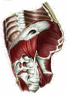
Abdominal muscles - 1866 illustration showing the structure of the muscles (brown) and tendons (white)
JAB4119475 Abdominal muscles - 1866 illustration showing the structure of the muscles (brown) and tendons (white) found in the front of the human abdomen - Region abdominale anterieure - Muscle

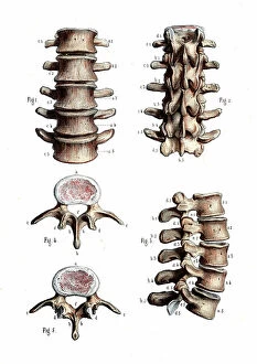
Background imageAnatomist Collection: Lumbar spine anatomy - 1866 anatomical illustration of the lumbar spine (lower back)

Lumbar spine anatomy - 1866 anatomical illustration of the lumbar spine (lower back) - Lumbar vertebrae
JAB4119471 Lumbar spine anatomy - 1866 anatomical illustration of the lumbar spine (lower back) - Lumbar vertebrae, vertebral column - Osteology, locomotion

Background imageAnatomist Collection: Chest and shoulder muscles - 1866 anatomical illustration showing the structure of the muscles

Chest and shoulder muscles - 1866 anatomical illustration showing the structure of the muscles (brown)
JAB4119460 Chest and shoulder muscles - 1866 anatomical illustration showing the structure of the muscles (brown) and tendons (white) of the rear of the upper back

Background imageAnatomist Collection: Foot anatomy - 1866 anatomical illustration showing the muscles (brown)

Foot anatomy - 1866 anatomical illustration showing the muscles (brown), tendons (white) and bones (beige)
JAB4119455 Foot anatomy - 1866 anatomical illustration showing the muscles (brown), tendons (white) and bones (beige) of the human foot - Plantaire region- Anatomy - Locomotion apparatus, myology

Background imageAnatomist Collection: Human hand - 1866 anatomical illustration showing the muscles (brown) and tendons (white)

Human hand - 1866 anatomical illustration showing the muscles (brown) and tendons (white)
JAB4119450 Human hand - 1866 anatomical illustration showing the muscles (brown) and tendons (white) of the human hand - Region palmar- Anatomy - Fig 1 Cubital anterieur Fig 2 cubital anterieur et

Background imageAnatomist Collection: Leg joints - 1866 anatomical illustration of the muscles (brown), tendons (white) and bones (beige)

Leg joints - 1866 anatomical illustration of the muscles (brown), tendons (white) and bones (beige)
JAB4119440 Leg joints - 1866 anatomical illustration of the muscles (brown), tendons (white) and bones (beige) of the human hip (top) and knee (bottom) joints

Background imageAnatomist Collection: Leg joints - 1866 anatomical illustration of the muscles (brown), tendons (white) and bones (beige)

Leg joints - 1866 anatomical illustration of the muscles (brown), tendons (white) and bones (beige)
JAB4119439 Leg joints - 1866 anatomical illustration of the muscles (brown), tendons (white) and bones (beige) of the human hip (top) and knee (bottom) joints

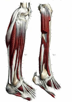
Background imageAnatomist Collection: Leg anatomy - 1866 anatomical illustration showing the muscles (brown)

Leg anatomy - 1866 anatomical illustration showing the muscles (brown), tendons (white) and bones (beige)
JAB4119436 Leg anatomy - 1866 anatomical illustration showing the muscles (brown), tendons (white) and bones (beige) of the human lower leg

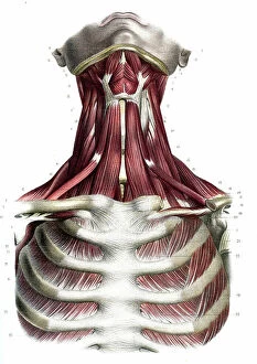
Background imageAnatomist Collection: Chest and neck muscles - 1866 anatomical illustration showing the structure of the muscles (brown)

Chest and neck muscles - 1866 anatomical illustration showing the structure of the muscles (brown) and tendons (white)
JAB4119434 Chest and neck muscles - 1866 anatomical illustration showing the structure of the muscles (brown) and tendons (white) found in the rear of the human head (top) and neck (upper centre)

Background imageAnatomist Collection: Neck muscles - 1866 illustration showing the structure of the muscles (brown) and tendons (white)

Neck muscles - 1866 illustration showing the structure of the muscles (brown) and tendons (white)
JAB4119427 Neck muscles - 1866 illustration showing the structure of the muscles (brown) and tendons (white) found in the exterior (left) and interior (right) of the human neck

Background imageAnatomist Collection: Chest and neck muscles - 1866 illustration showing the structure of the muscles (brown)

Chest and neck muscles - 1866 illustration showing the structure of the muscles (brown) and tendons (white)
JAB4119416 Chest and neck muscles - 1866 illustration showing the structure of the muscles (brown) and tendons (white) found in the human neck (upper centre) and chest (bottom)

Background imageAnatomist Collection: Human skull - 1866 illustration showing the anatomy of the human skull - La head - Descriptive

Human skull - 1866 illustration showing the anatomy of the human skull - La head - Descriptive anatomy - Fig 1
JAB4119396 Human skull - 1866 illustration showing the anatomy of the human skull - La head - Descriptive anatomy - Fig 1: Region laterale, Fig 2: Region superieure

Background imageAnatomist Collection: Leg joints - 1866 anatomical illustration showing the tendons (white) and bones (beige)

Leg joints - 1866 anatomical illustration showing the tendons (white) and bones (beige) of the human hip (top)
JAB4119386 Leg joints - 1866 anatomical illustration showing the tendons (white) and bones (beige) of the human hip (top) and knee (bottom) joints - Pelvis bone joints - Fig 1

Background imageAnatomist Collection: Knee and ankle joints - 1866 anatomical illustration showing the tendons (white) and bones (beige)

Knee and ankle joints - 1866 anatomical illustration showing the tendons (white) and bones (beige)
JAB4119379 Knee and ankle joints - 1866 anatomical illustration showing the tendons (white) and bones (beige) that make up the human knee and ankle joints- Fig 1 and 2

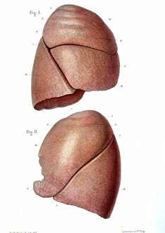
Background imageAnatomist Collection: Lung anatomy - 1866 illustration showing the external structure of the left (top) and right (bottom)

Lung anatomy - 1866 illustration showing the external structure of the left (top) and right (bottom)
JAB4119370 Lung anatomy - 1866 illustration showing the external structure of the left (top) and right (bottom) lungs - Lung - Descriptive anatomy - Fig 1: right lung, outer face, Fig 2: left lung

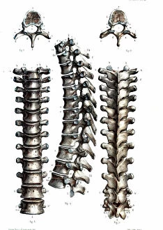
Background imageAnatomist Collection: Thoracic spine anatomy - 1866 anatomical illustration of the thoracic spine

Thoracic spine anatomy - 1866 anatomical illustration of the thoracic spine
JAB4119368 Thoracic spine anatomy - 1866 anatomical illustration of the thoracic spine, showing the individual vertebrae - Vertebral column - Dorsal vertebrae, osteology, locomotion

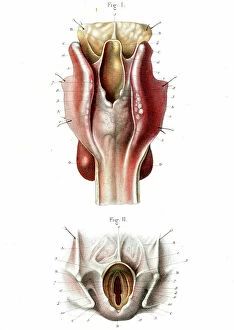
Background imageAnatomist Collection: Human throat 1866 cutaway illustration showing the anatomy of the throat LE LARYNX

Human throat 1866 cutaway illustration showing the anatomy of the throat LE LARYNX
JAB4119196 Human throat 1866 cutaway illustration showing the anatomy of the throat LE LARYNX, posterieure face - Fig 1: Pharynx and esophagus, Fig 2: Opening of the larynx 5 sup-glottic part

Background imageAnatomist Collection: Voice box anatomy 1866 anatomical illustration showing the muscles (brown)

Voice box anatomy 1866 anatomical illustration showing the muscles (brown) and external (Figs 1 and 2)
JAB4119181 Voice box anatomy 1866 anatomical illustration showing the muscles (brown) and external (Figs 1 and 2) and internal structure (Fig 3) of the larynx (voice box) Muscles du larynx - Fig 1

Background imageAnatomist Collection: Diaphragm 1866

Diaphragm 1866
JAB4119165 Diaphragm 1866; (add.info.: Diaphragm 1866 illustration of a section through the abdomen and chest, showing the diaphragm (arched)

Background imageAnatomist Collection: Menstrual cycle anatomy 1866

Menstrual cycle anatomy 1866
JAB4119155 Menstrual cycle anatomy 1866; (add.info.: Menstrual cycle anatomy 1866 illustrations showing the internal anatomy of a human ovary containing Graafian (ovarian) follicles (Fig 1)

Background imageAnatomist Collection: Male pelvic blood supply 1866 illustration showing the major veins (blue) and arteries (red)

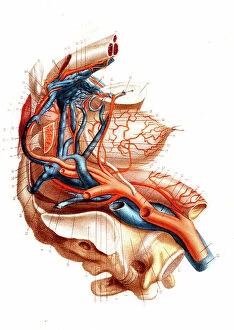
Male pelvic blood supply 1866 illustration showing the major veins (blue) and arteries (red)
JAB4119143 Male pelvic blood supply 1866 illustration showing the major veins (blue) and arteries (red) of the male pelvis These include the inferior vena cava, common iliac vein

Background imageAnatomist Collection: BARTHOLIN, Thomas (1616-1680). Danish physician, mathematician, and theologian

BARTHOLIN, Thomas (1616-1680). Danish physician, mathematician, and theologian
CUL4875648 BARTHOLIN, Thomas (1616-1680). Danish physician, mathematician, and theologian. He is best known for his work in the discovery of the lymphatic system in humans

Background imageAnatomist Collection: study of a body, hand, copperplate engraving, about 1700

study of a body, hand, copperplate engraving, about 1700
UTD4921972 study of a body, hand, copperplate engraving, about 1700; (add.info.: study of a body, hand, copperplate engraving)

Background imageAnatomist Collection: The eye , copperplate engraving, about 1700

The eye , copperplate engraving, about 1700
UTD4921822 The eye , copperplate engraving, about 1700; (add.info.: The eye , copperplate engraving, about 1700 ©HistAuge/United Archives/Leemage); United Archives/HistAuge.

Background imageAnatomist Collection: Anatomy of the human arterial system in the legs. Handcoloured lithograph by St

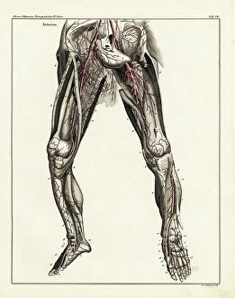
Anatomy of the human arterial system in the legs. Handcoloured lithograph by St
FLO4684812 Anatomy of the human arterial system in the legs. Handcoloured lithograph by St. Schillinger from Lorenz Oken's Universal Natural History, Allgemeine Naturgeschichte fur alle Stande


All Professionally Made to Order for Quick Shipping

Anatomists have played a crucial role in unraveling the mysteries of the human body throughout history. One such pioneer was Robert Burton, whose extensive research and writings on anatomy left an indelible mark on the field. Another notable figure was EUSTACHIO Bartolomeo, an Italian anatomist who made significant contributions during the 16th century. Intriguingly, a lithograph titled "Odin questioning Mimir" captures the essence of curiosity that drives anatomists to seek knowledge. This thirst for understanding is exemplified by Sir Richard Owen, a renowned British anatomist from the 19th century whose work continues to inspire modern scientists. Richard Owen's dedication to his craft is evident in various depictions of him throughout history. In one caricature from Vanity Fair in 1873, aptly titled "Old bones, " he is portrayed as a distinguished scholar with wisdom etched onto his face. An image from his study at BMNH in 1883 showcases his meticulous approach towards dissecting and studying specimens. The intricate details captured in engravings like "The Sacred Heart of Jesus" or "The pollen of plants" demonstrate how anatomists explore not only human anatomy but also delve into other aspects of nature's design. Richard Owen himself had a profound impact on paleontology, as depicted in C016 / 5008 - an image that pays homage to his groundbreaking discoveries. Throughout history, heart anatomists have held a special place within this scientific realm. Figures like Guillaume Dupuytren and William Hunter dedicated their lives to unraveling the complexities of this vital organ. Their contributions paved the way for advancements in cardiac medicine that continue to save lives today. From ancient times until now, these brilliant minds have shaped our understanding of ourselves and our world through their tireless pursuit of knowledge about our bodies' inner workings. The legacy they leave behind serves as an inspiration for future generations aspiring to unravel the secrets of human anatomy.