Fraud Blocker Skip to main content

Diseased Collection (page 4)

"Diseased: A Glimpse into the World of Health Challenges" Bunions and X-ray: Unveiling the Painful Reality of Foot Deformities

Background imageDiseased Collection: Healthy and diseased organs, artwork

Healthy and diseased organs, artwork
Healthy and diseased female organs, computer artwork

Background imageDiseased Collection: Immunotherapy, artwork

Immunotherapy, artwork
Immunotherapy. Computer artwork of immunotherapy being used to destroy a tumour (right). Immunotherapy uses the bodys immune system to fight a disease

Background imageDiseased Collection: Effect of Parkinsons disease, artwork

Effect of Parkinsons disease, artwork
Effect of Parkinsons disease. Computer artwork showing the neural pathways within the brain that are affected by a lack of dopamine, caused by the onset of Parkinsons disease

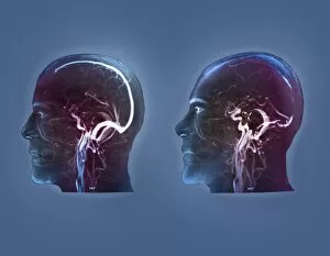
Background imageDiseased Collection: Thrombophlebitis in the brain, MRI scans

Thrombophlebitis in the brain, MRI scans
Thrombophlebitis in the brain. Coloured magnetic resonance imaging (MRI) angiograms comparing the blood vessels (white) of the brain and neck in a healthy 43-year-old (left)

Background imageDiseased Collection: Thrombophlebitis in the brain, 3D CT scan

Thrombophlebitis in the brain, 3D CT scan
Thrombophlebitis in the brain. Coloured 3D computed tomography (CT) angiogram of the blood vessels (blue) in the brain of a 48-year-old with thrombophlebitis of the superior sagittal sinus

Background imageDiseased Collection: Gorhams disease, 3D CT scan

Gorhams disease, 3D CT scan
Gorhams disease. Coloured 3D computed tomography (CT) scan of the rear of the skull of a 47-year-old patient, showing damage (centre) caused by osteolysis (bone resorption and replacement)

Background imageDiseased Collection: Nerve cell degeneration, artwork

Nerve cell degeneration, artwork
Nerve cell degeneration. Computer artwork showing a collection of nerve cells (neurons) that have become damaged

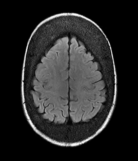
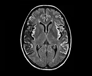
Background imageDiseased Collection: Thickened skull, MRI scan

Thickened skull, MRI scan
Thickened skull. Magnetic resonance imaging (MRI) scan of an axial section through the head of a 22-year-old patient, showing thickening of the trabecular (spongy) bone of the skull

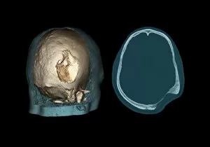
Background imageDiseased Collection: Gorhams disease, CT scans

Gorhams disease, CT scans
Gorhams disease. Coloured 3D (left) and 2D (right) computed tomography (CT) scans of the skull of a 47-year-old patient, showing damage (centre) caused by osteolysis (bone resorption and replacement)

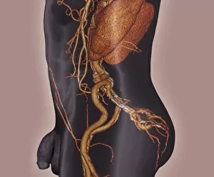
Background imageDiseased Collection: Atherosclerosis, 3D CT scan

Atherosclerosis, 3D CT scan
Atherosclerosis. 3D computed tomography (CT) angiogram of the main blood vessels of the lower body a 58-year-old man, showing numerous atheromatous plaques (light, centre) on the vessel walls

Background imageDiseased Collection: Substantia nigra, artwork

Substantia nigra, artwork
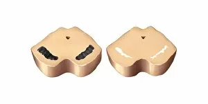
Substantia nigra. Computer artwork showing a healthy substantia nigra (left) and degenerated substantia nigra (right) from human brains

Background imageDiseased Collection: Pseudomonas aeruginosa bacterium, SEM C016 / 9442

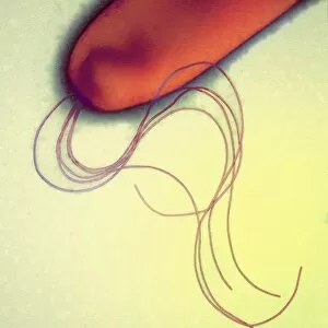
Pseudomonas aeruginosa bacterium, SEM C016 / 9442
Pseudomonas aeruginosa bacterium, coloured transmission electron micrograph (TEM). P. aeruginosa is a Gram-negative rod-shaped bacterium found in soil and water

Background imageDiseased Collection: Mycobacterium fortuitum bacteria C016 / 9424

Mycobacterium fortuitum bacteria C016 / 9424
Mycobacterium fortuitum bacteria, coloured scanning electron micrograph (SEM). These bacteria (blue) are a non-tuberculosis strain of Mycobacterium

Background imageDiseased Collection: Lassa virus particles, TEM C016 / 9409

Lassa virus particles, TEM C016 / 9409
Lassa virus particles. Transmission electron micrograph (TEM) of Lassa virus particles (virions, blue) amongst cell debris. This Arenavirus is the cause of Lassa fever

Background imageDiseased Collection: Orientia tsutsugamushi bacterium C016 / 9431

Orientia tsutsugamushi bacterium C016 / 9431
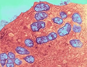
Orientia tsutsugamushi bacterium. Transmission electron micrograph (TEM) of a section through an Orientia tsutsugamushi bacterium. O

Background imageDiseased Collection: Pseudomonas aeruginosa bacterium, SEM C016 / 9441

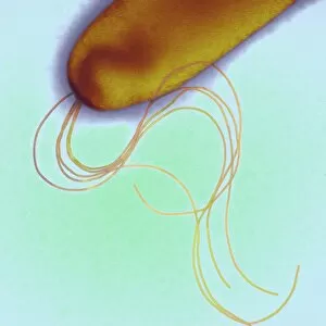
Pseudomonas aeruginosa bacterium, SEM C016 / 9441
Pseudomonas aeruginosa bacterium, coloured transmission electron micrograph (TEM). P. aeruginosa is a Gram-negative rod-shaped bacterium found in soil and water

Background imageDiseased Collection: Acute haemorrhagic conjunctivitis, TEM C016 / 9387

Acute haemorrhagic conjunctivitis, TEM C016 / 9387
Acute haemorrhagic conjunctivitis. Transmission electron micrograph (TEM) of a sample of tissue from the conjunctiva of a human eye that is infected by enterovirus particles (purple)

Background imageDiseased Collection: Eastern equine encephalitis, TEM C016 / 9390

Eastern equine encephalitis, TEM C016 / 9390
Eastern equine encephalitis. Transmission electron micrograph (TEM) of a section through tissue from a human central nervous system (CNS)

Background imageDiseased Collection: Colorado tick fever virus infection, TEM C016 / 9384

Colorado tick fever virus infection, TEM C016 / 9384
Colorado tick fever virus infection. Transmission electron micrograph (TEM) of human tissue infected by Colorado tick fever virus (CTFV) particles (virions, dark circles)

Background imageDiseased Collection: Syphilis bacterium, TEM C016 / 9461

Syphilis bacterium, TEM C016 / 9461
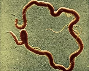
Syphilis bacteria. Transmission electron micrograph (TEM) of a Treponema pallidum spirochaete bacterium. T. pallidum is the bacteria that is responsible for the sexually transmitted infection syphilis

Background imageDiseased Collection: Swine flu virus particles, TEM C016 / 9399

Swine flu virus particles, TEM C016 / 9399
Swine flu virus particles. Transmission electron micrograph (TEM) of a section through H3N2 influenza A virus particles (virions)

Background imageDiseased Collection: Swine flu virus particles, TEM C016 / 9406

Swine flu virus particles, TEM C016 / 9406
Influenza virus particles. Transmission electron micrograph (TEM) of influenza (flu) virus particles (virions). Each virion consists of ribonucleic acid (RNA, dark patches)

Background imageDiseased Collection: Syphilis bacterium, TEM C016 / 9462

Syphilis bacterium, TEM C016 / 9462
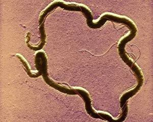
Syphilis bacteria. Transmission electron micrograph (TEM) of a Treponema pallidum spirochaete bacterium. T. pallidum is the bacteria that is responsible for the sexually transmitted infection known

Background imageDiseased Collection: HIV virus particles, TEM C016 / 9404

HIV virus particles, TEM C016 / 9404
HIV virus particles. Transmission electron micrograph (TEM) of a section through human immunodeficiency virus (HIV) particles (virions, round)

Background imageDiseased Collection: Mycobacterium fortuitum bacteria C016 / 9423

Mycobacterium fortuitum bacteria C016 / 9423
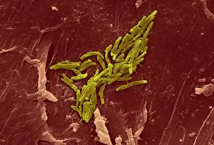
Mycobacterium fortuitum bacteria, coloured scanning electron micrograph (SEM). These bacteria (yellow) are a non-tuberculosis strain of Mycobacterium

Background imageDiseased Collection: Gonorrhoea bacteria, TEM C016 / 9425

Gonorrhoea bacteria, TEM C016 / 9425
Gonorrhoea bacteria. Transmission electron micrograph (TEM) of a diplococcal pair of Neisseria gonorrhoeae bacteria. N. gonorrhoeae is the bacteria that is responsible for the sexually transmitted

Background imageDiseased Collection: HIV virus particles, TEM C016 / 9405

HIV virus particles, TEM C016 / 9405
HIV virus particles. Transmission electron micrograph (TEM) of a section through human immunodeficiency virus (HIV) particles (virions, round)

Background imageDiseased Collection: Swine flu virus particles, TEM C016 / 9400

Swine flu virus particles, TEM C016 / 9400
Swine flu virus particles. Transmission electron micrograph (TEM) of a section through H3N2 influenza A virus particles (virions)

Background imageDiseased Collection: Orientia tsutsugamushi bacteria infection C016 / 9429

Orientia tsutsugamushi bacteria infection C016 / 9429
Orientia tsutsugamushi bacteria infection. Transmission electron micrograph (TEM) of a section through a capillary (red) infected with Orientia tsutsugamushi bacteria (blue). O

Background imageDiseased Collection: Lassa virus particles, TEM C016 / 9408

Lassa virus particles, TEM C016 / 9408
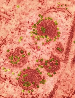
Lassa virus particles. Transmission electron micrograph (TEM) of Lassa virus particles (virions, green) amongst cell debris. This Arenavirus is the cause of Lassa fever

Background imageDiseased Collection: Orientia tsutsugamushi bacterium C016 / 9430

Orientia tsutsugamushi bacterium C016 / 9430
Orientia tsutsugamushi bacterium. Transmission electron micrograph (TEM) of a section through an Orientia tsutsugamushi bacterium. O

Background imageDiseased Collection: Ganjam virus infection, TEM C016 / 9396

Ganjam virus infection, TEM C016 / 9396
Ganjam virus infection. Transmission electron micrograph (TEM) of a section through human tissue infected with ganjam virus particles (virions, blue and yellow)

Background imageDiseased Collection: Swine flu virus particles, TEM C016 / 9407

Swine flu virus particles, TEM C016 / 9407
Influenza virus particles. Transmission electron micrograph (TEM) of influenza (flu) virus particles (virions). Each virion consists of ribonucleic acid (RNA, dark patches)

Background imageDiseased Collection: Flu virus, TEM

Flu virus, TEM
Flu virus. Transmission electron micrograph (TEM) of a section through influenza (flu) virus particles (virions, purple) budding from a host cell. This is the final stage in viral replication

Background imageDiseased Collection: Ductal breast cancer, TEM

Ductal breast cancer, TEM
Ductal breast cancer. Transmission electron micrograph (TEM) of a section through breast tissue, showing malignant (cancerous) epithelial cells arising from the milk ducts of the breast

Background imageDiseased Collection: Colorectal cancer cell, SEM

Colorectal cancer cell, SEM
Colorectal cancer cell. Coloured scanning electron micrograph (SEM) of a cancer cell from the human colon (large intestine). Cancer of the colon is also known as colorectal cancer

Background imageDiseased Collection: Creutzfeldt-Jakob disease, MRI scan

Creutzfeldt-Jakob disease, MRI scan
Creutzfeldt-Jakob disease. Coloured magnetic resonance imaging (MRI) scan of an axial section through the brain of a 35-year-old patient

Background imageDiseased Collection: Kidney tumour, CT scan

Kidney tumour, CT scan
Kidney tumour. Coloured computed tomography (CT) scan of a section through the abdomen of a 62-year-old patient, showing necrosis (death) of tissue in the right kidney

Background imageDiseased Collection: Light-reactive dental filling, artwork

Light-reactive dental filling, artwork
Light-reactive dental filling. Computer artwork showing a blue light (top left) being used to harden a composite dental filling in a tooth (lower right)

Background imageDiseased Collection: Leukaemia cell, SEM

Leukaemia cell, SEM
Leukaemia cell. Coloured scanning electron micrograph (SEM) of a leukaemic (cancerous) lymphocyte white blood cell (green), amongst normal red blood cells (erythrocytes, red)

Background imageDiseased Collection: Root canal dental treatment, artwork

Root canal dental treatment, artwork
Root canal dental treatment. Computer artwork showing a drill (top left) being used to clear out the root canals (blue) of a decayed tooth

Background imageDiseased Collection: Oncogenes, artwork C016 / 6262

Oncogenes, artwork C016 / 6262
Oncogenes. Computer artwork comparing the DNA (deoxyribonucleic acid, coiled strand) of a normal cell (top) with that of a cancer cell (bottom)

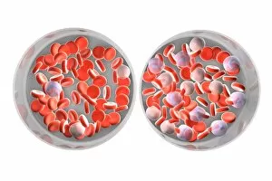
Background imageDiseased Collection: Normal and leukaemic blood, artwork C016 / 6261

Normal and leukaemic blood, artwork C016 / 6261
Normal and leukaemic blood. Computer artwork comparing the appearance of normal blood (left) and that affected by leukaemia (right)

Background imageDiseased Collection: Effect of oxygen supply on tumour growth C016 / 6256

Effect of oxygen supply on tumour growth C016 / 6256
Effect of oxygen supply on tumour growth. Top: a small blood supply (tubes) means a low level of oxygen is available and the tumour (round) grows slowly

Background imageDiseased Collection: Cirrhosis of liver, light micrograph C016 / 0530

Cirrhosis of liver, light micrograph C016 / 0530
Cirrhosis of liver. Light micrograph of a section through liver tissue, damaged by cirrhosis. Cirrhosis occurs as a result of a range of factors causing damage to liver function

Background imageDiseased Collection: Bowel cancer, light micrograph C016 / 0511

Bowel cancer, light micrograph C016 / 0511
Bowel cancer. Light micrograph of a section through a carcinoid tumour in the small bowel. The tumour has invaded the submucosa of the bowel

Background imageDiseased Collection: Cirrhosis of liver, light micrograph C016 / 0529

Cirrhosis of liver, light micrograph C016 / 0529
Cirrhosis of liver. Light micrograph of a section through liver tissue, damaged by cirrhosis. Cirrhosis occurs as a result of a range of factors causing damage to liver function

Background imageDiseased Collection: Gingivitis, SEM C014 / 1443

Gingivitis, SEM C014 / 1443
Gingivitis. Coloured scanning electron micrograph (SEM) of a large number of bacteria (round) in the gingiva (gums) of a human mouth


All Professionally Made to Order for Quick Shipping

"Diseased: A Glimpse into the World of Health Challenges" Bunions and X-ray: Unveiling the Painful Reality of Foot Deformities. Coral Spot Fungus on Sycamore Twig: Nature's Intricate Battle with Disease. Tension Pneumothorax Revealed: When Every Breath Becomes a Struggle. Bacterial Meningitis Unmasked: Peering into the Depths of an Invisible Threat. The Diseased Heart: An Organ in Need of Healing and Care. Malaria Parasites Under TEM Microscope: Understanding the Silent Invaders Within Us. Historic Advertisement for Migranin: Tracing Headache Relief Through Time and Culture. Consultation Hour at Sebastianeum Spa Hotel, Bavaria, 1880s: Seeking Remedies Amidst Serenity. The Spa Doctor in Thuringia, Germany: Bridging Medicine and Wellness in Centuries Past. At the Sickbed - A Mother's Vigilance Over Her Ill Child, France 1881: Love Transcends Disease's Grip. Bei der Dorfsybille - Prophetess from Germany 1881: Ancient Wisdom Meets Modern Maladies Caring for Casualties at Berlin Ambulance Station, 1895 Germany .