
Diseased Collection (page 7)
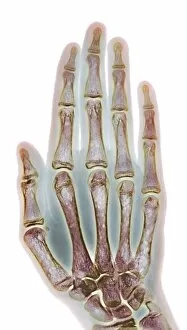
"Diseased: A Glimpse into the World of Health Challenges" Bunions and X-ray: Unveiling the Painful Reality of Foot Deformities


All Professionally Made to Order for Quick Shipping
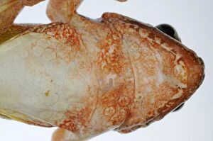
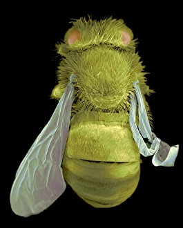
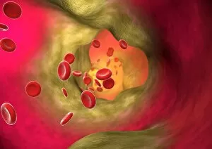
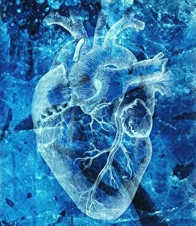
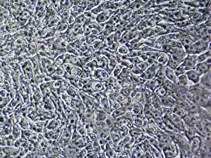
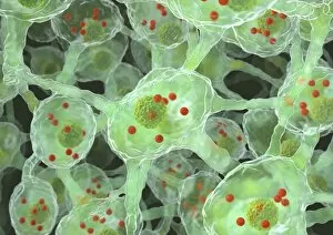
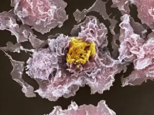
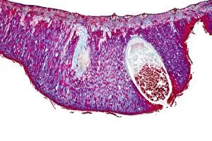
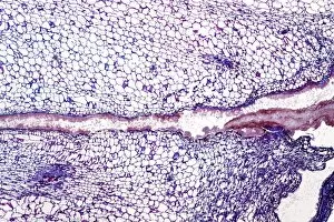
"Diseased: A Glimpse into the World of Health Challenges" Bunions and X-ray: Unveiling the Painful Reality of Foot Deformities. Coral Spot Fungus on Sycamore Twig: Nature's Intricate Battle with Disease. Tension Pneumothorax Revealed: When Every Breath Becomes a Struggle. Bacterial Meningitis Unmasked: Peering into the Depths of an Invisible Threat. The Diseased Heart: An Organ in Need of Healing and Care. Malaria Parasites Under TEM Microscope: Understanding the Silent Invaders Within Us. Historic Advertisement for Migranin: Tracing Headache Relief Through Time and Culture. Consultation Hour at Sebastianeum Spa Hotel, Bavaria, 1880s: Seeking Remedies Amidst Serenity. The Spa Doctor in Thuringia, Germany: Bridging Medicine and Wellness in Centuries Past. At the Sickbed - A Mother's Vigilance Over Her Ill Child, France 1881: Love Transcends Disease's Grip. Bei der Dorfsybille - Prophetess from Germany 1881: Ancient Wisdom Meets Modern Maladies Caring for Casualties at Berlin Ambulance Station, 1895 Germany .