Fraud Blocker Skip to main content

Diseased Collection (page 8)

"Diseased: A Glimpse into the World of Health Challenges" Bunions and X-ray: Unveiling the Painful Reality of Foot Deformities

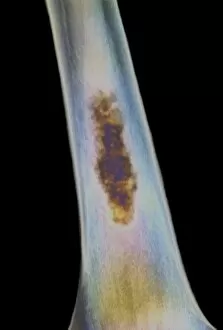
Background imageDiseased Collection: Bone death, X-ray

Bone death, X-ray
Bone death. Coloured X-ray of the shaft of a human long bone showing tissue death (osteonecrosis, brown). This may be caused by a blood clot or embolus that interrupts the blood supply

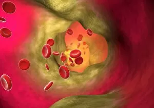
Background imageDiseased Collection: Blocked artery

Blocked artery
Artery disease. Computer artwork of blood cells (round) in an artery affected by atherosclerosis. This is a narrowing of an artery due to fatty deposits (yellow) on its inner walls

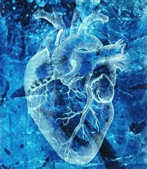
Background imageDiseased Collection: Unhealthy heart

Unhealthy heart, conceptual image. The main blood vessels of the heart include the vena cava (upper left), where blood arrives form the body

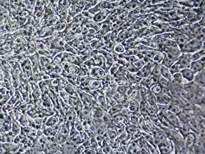
Background imageDiseased Collection: Cystic fibrosis stem cells

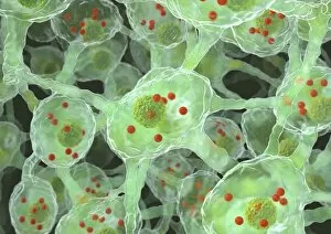
Cystic fibrosis stem cells. Light micrograph of human embryonic stem cells containing the mutation for cystic fibrosis (CF). Stem cells are able to differentiate into other cell types (pluripotent)

Background imageDiseased Collection: Calf

Calf. Blurred head of a young dairy cow (Bos taurus). This image could be used to illustrate " mad cow disease" (bovine spongiform encephalopathy, BSE)

Background imageDiseased Collection: Parkinsons disease nerve cells, artwork

Parkinsons disease nerve cells, artwork
Parkinsons disease nerve cells. Computer artwork of human nerve cells (neurons, green) affected by Lewy bodies (orange) in the brain of someone with Parkinsons disease

Background imageDiseased Collection: TB bacteria infecting macrophages, SEM

TB bacteria infecting macrophages, SEM
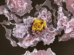
TB bacteria infecting macrophages. Coloured scanning electron micrograph (SEM) of Mycobacterium tuberculosis bacteria (orange) invading macrophage white blood cells (pink)

Background imageDiseased Collection: Sexually transmitted infection, artwork

Sexually transmitted infection, artwork
Sexually transmitted infection, conceptual computer artwork

Background imageDiseased Collection: Pear rust fungus, light micrograph

Pear rust fungus, light micrograph
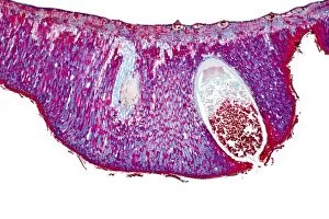
Pear rust fungus. Light micrograph of a section through tissue from a juniper tree infected with pear rust fungus (Gymnosporangium sabinae, lower right)

Background imageDiseased Collection: Grey mould fungus, light micrograph

Grey mould fungus, light micrograph
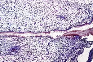
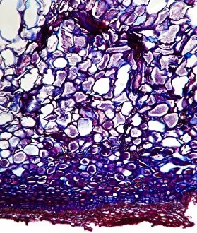
Grey mould fungus. Light micrograph of a section through onion tissue infected with grey mould fungus (Botrytis allii). Botrytis is a parasitic saprophyte mould that grows on wounded plants

Background imageDiseased Collection: Corn smut infection, light micrograph

Corn smut infection, light micrograph
Corn smut infection. Light micrograph of a section through tissue from a corn (Zea mays) plant infected with corn smut fungus (Ustilago maydis)

Background imageDiseased Collection: Bramble rust fungus spores

Bramble rust fungus spores. Light micrograph of teliospores from the bramble rust fungus (Phragmidium violaceum). Magnification: x37 when printed 10 centimetres wide

Background imageDiseased Collection: Rose mildew fungus, light micrograph

Rose mildew fungus, light micrograph
Rose mildew fungus. Light micrograph of a section through tissue from a rose plant infected with rose mildew fungus (Erysiphe pannosa)

Background imageDiseased Collection: Pear scab infection, light micrograph

Pear scab infection, light micrograph
Pear scab infection. Light micrograph of a section through a pear (Pyrus sp.) infected with the Venturia pirinum fungus. This fungus causes scab, or black spot, of the pear fruit

Background imageDiseased Collection: Black scab infection, light micrograph

Black scab infection, light micrograph
Black scab infection. Light micrograph of a section through a potato infected with black scab (Synchytrium endobioticum) fungus

Background imageDiseased Collection: White rust fungus on a leaf

White rust fungus on a leaf. Light micrograph of a section through a plant leaf infected with white rust fungus (Albugo candida)

Background imageDiseased Collection: Powdery scab infection, light micrograph

Powdery scab infection, light micrograph
Powdery scab infection. Light micrograph of a section through a potato infected with powdery scab (Spongospora subterranea), showing pustules and spores. S

Background imageDiseased Collection: Plum pocket infection, light micrograph

Plum pocket infection, light micrograph
Plum pocket infection. Light micrograph of a section through a plum gall caused by the plum pocket fungus (Taphrina pruni)

Background imageDiseased Collection: Rust fungus in a leaf, light micrograph

Rust fungus in a leaf, light micrograph
Rust fungus in a leaf. Light micrograph of a section through a wheat leaf infected with the parasitic Puccinia graminis rust

Background imageDiseased Collection: Penicillium mould, light micrograph

Penicillium mould, light micrograph
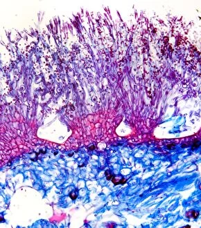
Penicillium mould. Light micrograph of a section through an orange rind infected with Penicillium sp. blue mould fungus, showing fungal hyphae and conidiophores (pink and purple) with spores (red)

Background imageDiseased Collection: Brown mould fungus, light micrograph

Brown mould fungus, light micrograph
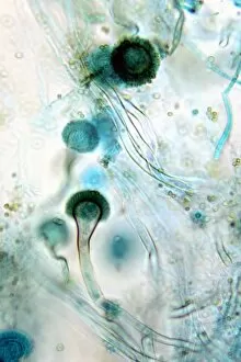
Brown mould fungus. Light micrograph of a section through decaying plant tissue infected with brown mould fungus (Aspergillus glaucum)

Background imageDiseased Collection: Downy mildew infection, light micrograph

Downy mildew infection, light micrograph
Downy mildew infection. Light micrograph of a section through a grapevine (Vitis sp.) leaf infected with downy mildew (Plasmopara viticola)

Background imageDiseased Collection: Hollyhock rust fungus in a leaf

Hollyhock rust fungus in a leaf. Light micrograph of a section through a hollyhock (Alcea sp.) leaf infected with the parasitic Puccinia malvacearum rust, showing the teliospores (spores)

Background imageDiseased Collection: Clubroot infection, light micrograph

Clubroot infection, light micrograph
Clubroot infection. Light micrograph a section through a cabbage root infected with clubroot (Plasmodiophora brassicae). Plasmodiophora is a parasitic protist that infects the roots of cabbages

Background imageDiseased Collection: Arthritic hands

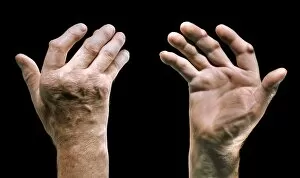
Arthritic hands
MODEL RELEASED. Arthritic hands. Hands of a person that have become deformed due to arthritis. Arthritis is a degenerative condition that affects the joints causing inflammation and pain

Background imageDiseased Collection: Balloon angioplasty, artwork

Balloon angioplasty, artwork
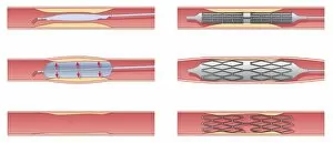
Balloon angioplasty. Artwork showing balloon angioplasty treatment for stenosis (narrowing) of a coronary artery. A balloon catheter (blue)

Background imageDiseased Collection: Angioplasty, artwork

Angioplasty, artwork
Angioplasty. Artwork showing two treatments for stenosis (narrowing) of arteries. At left is balloon angioplasty, where a balloon catheter is inserted into the affected artery

Background imageDiseased Collection: Narrowed blood vessel, artwork

Narrowed blood vessel, artwork
Narrowed blood vessel. Cut-away artwork showing a blood vessel that is partially blocked by atheroma. Atheroma is the deposition of fatty material on the inner walls of a blood vessel

Background imageDiseased Collection: Bone death, artwork

Bone death, artwork
Bone death. Computer artwork of a human hip and knee joint affected by osteonecrosis - death of the bone tissue. This condition is caused by an impaired blood supply to the bone cells

Background imageDiseased Collection: Childhood rickets, X-ray

Childhood rickets, X-ray
Childhood rickets. Coloured frontal X-ray of the legs of a 3-year-old child with rickets. Rickets (Osteomalacia) occurs predominantly in children

Background imageDiseased Collection: Tooth decay, X-ray

Tooth decay, X-ray
Tooth decay. Coloured X-ray of a profile view of a 22-year-old patients teeth, showing a cavity (decay) in the left lower molar (light grey, centre left)

Background imageDiseased Collection: Chronic liver disease, CT scan

Chronic liver disease, CT scan
Chronic liver disease. Coloured computed tomography (CT) scan of an axial section through the abdominopelvic region of a 58-year-old patient with a history of hepatitis C

Background imageDiseased Collection: Slipped disc, MRI scan

Slipped disc, MRI scan
Slipped disc, coloured magnetic resonance imaging (MRI) scan. Profile view of the spine of a 59-year-old patient with severe rheumatoid arthritis and a herniated (slipped) disc (red)

Background imageDiseased Collection: Spine in Hurler Syndrome, X-ray

Spine in Hurler Syndrome, X-ray
Spine in Hurler Syndrome. Coloured X-ray of the spine of a 26-month-old child with Hurler syndrome (also called mucopolysaccharidosis type I, MPS I)

Background imageDiseased Collection: Osteoarthritis of the knee, artwork

Osteoarthritis of the knee, artwork
Osteoarthritis of the knee. Computer artwork showing a knee joint affected by osteoarthritis. Osteoarthritis is a degenerative disease that involves degradation of the bone

Background imageDiseased Collection: Parkinsons disease, artwork

Parkinsons disease, artwork
Parkinsons disease. Computer artwork of neurons (nerve cells, pink) containing Lewy bodies (green). Lewy bodies, which are deposits of protein

Background imageDiseased Collection: Oak spangle galls

Oak spangle galls
Common spangle galls of the gall wasp Neuroterus quercusbaccarum on underside of oak leaf. Dorset UK September 2009

Background imageDiseased Collection: Alder bead galls

Alder bead galls on a black alder (Alnus glutinosa) leaf. These galls are caused by the alder gall mite (Phytoptus laevis)

Background imageDiseased Collection: Aphid galls

Aphid galls
Apid galls on a Lombardy poplar (Populus nigra Italica) leaf. The two galls are caused by two different species of aphid. The gall on the leaf stalk is caused by Pemphigus spyrothecae

Background imageDiseased Collection: Knopper gall on acorn

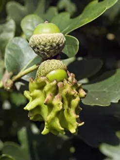
Knopper gall on acorn. Close-up of two acorns from an Pedunculate Oak (Quercus robur) tree. The lower one has been infected by the parasitic larva of a gall wasp (Andricus quercuscalicis)

Background imageDiseased Collection: Artichoke Galls on Pedunculate Oak

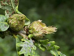
Artichoke Galls on Pedunculate Oak (Quercus rubus). Flower buds infected by the gall wasp Andricus fecundator. In spring the female gall wasp lays single eggs in buds of the Pedunculate oak

Background imageDiseased Collection: Spread of disease

Spread of disease. Conceptual computer artwork of Earth over-run with bacteria

Background imageDiseased Collection: Tar spot fungus on sycamore leaves

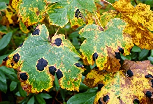
Tar spot fungus on sycamore leaves. Leaves of a sycamore (Acer pseudoplatanus) tree, affected by the fungal pathogen Rhytisma acerinum, which causes a disease known as tar spot

Background imageDiseased Collection: Palm seeds infected with pestalotia

Palm seeds infected with pestalotia
Palm seeds infected with the parasitic fungus Pestalotia sp. (black growths)

Background imageDiseased Collection: Red Fox - adult badly affected by mange - Wiltshire - England - UK

Red Fox - adult badly affected by mange - Wiltshire - England - UK
DK-362 Red Fox - adult badly affected by mange Wiltshire - England - UK Vulpes vulpes David Kilbey Please note that prints are for personal display purposes only and may not be reproduced in any way

Background imageDiseased Collection: Rabbit in late stages of Myxomatosis showing swelling around eyes, Cotswolds, UK

Rabbit in late stages of Myxomatosis showing swelling around eyes, Cotswolds, UK
MAB-89 Rabbit in late stages of Myxomatosis showing swelling around eyes Cotswolds, UK Mark Boulton Please note that prints are for personal display purposes only

Background imageDiseased Collection: Jesus Heals Multitude

Jesus Heals Multitude
Christ healeth a Multitude of diseased Persons at Genezareth

Background imageDiseased Collection: Sclerotium Clavus

Sclerotium Clavus
ERGOT of rye seed, a diseased transformation by a fungus, which can lead to poisoning by contaminated bread. Right : TILLETIA CARIES, another harmful growth


All Professionally Made to Order for Quick Shipping

"Diseased: A Glimpse into the World of Health Challenges" Bunions and X-ray: Unveiling the Painful Reality of Foot Deformities. Coral Spot Fungus on Sycamore Twig: Nature's Intricate Battle with Disease. Tension Pneumothorax Revealed: When Every Breath Becomes a Struggle. Bacterial Meningitis Unmasked: Peering into the Depths of an Invisible Threat. The Diseased Heart: An Organ in Need of Healing and Care. Malaria Parasites Under TEM Microscope: Understanding the Silent Invaders Within Us. Historic Advertisement for Migranin: Tracing Headache Relief Through Time and Culture. Consultation Hour at Sebastianeum Spa Hotel, Bavaria, 1880s: Seeking Remedies Amidst Serenity. The Spa Doctor in Thuringia, Germany: Bridging Medicine and Wellness in Centuries Past. At the Sickbed - A Mother's Vigilance Over Her Ill Child, France 1881: Love Transcends Disease's Grip. Bei der Dorfsybille - Prophetess from Germany 1881: Ancient Wisdom Meets Modern Maladies Caring for Casualties at Berlin Ambulance Station, 1895 Germany .