Fraud Blocker Skip to main content

Diseased Collection (page 5)

"Diseased: A Glimpse into the World of Health Challenges" Bunions and X-ray: Unveiling the Painful Reality of Foot Deformities

Background imageDiseased Collection: Colorectal cancer cell, SEM

Colorectal cancer cell, SEM
Colorectal cancer cell. Coloured scanning electron micrograph (SEM) of a cancer cell from the human colon (large intestine). Cancer of the colon is also known as colorectal cancer

Background imageDiseased Collection: Creutzfeldt-Jakob disease, MRI scan

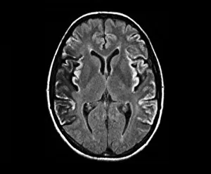
Creutzfeldt-Jakob disease, MRI scan
Creutzfeldt-Jakob disease. Coloured magnetic resonance imaging (MRI) scan of an axial section through the brain of a 35-year-old patient

Background imageDiseased Collection: Kidney tumour, CT scan

Kidney tumour, CT scan
Kidney tumour. Coloured computed tomography (CT) scan of a section through the abdomen of a 62-year-old patient, showing necrosis (death) of tissue in the right kidney

Background imageDiseased Collection: Light-reactive dental filling, artwork

Light-reactive dental filling, artwork
Light-reactive dental filling. Computer artwork showing a blue light (top left) being used to harden a composite dental filling in a tooth (lower right)

Background imageDiseased Collection: Leukaemia cell, SEM

Leukaemia cell, SEM
Leukaemia cell. Coloured scanning electron micrograph (SEM) of a leukaemic (cancerous) lymphocyte white blood cell (green), amongst normal red blood cells (erythrocytes, red)

Background imageDiseased Collection: Root canal dental treatment, artwork

Root canal dental treatment, artwork
Root canal dental treatment. Computer artwork showing a drill (top left) being used to clear out the root canals (blue) of a decayed tooth

Background imageDiseased Collection: Oncogenes, artwork C016 / 6262

Oncogenes, artwork C016 / 6262
Oncogenes. Computer artwork comparing the DNA (deoxyribonucleic acid, coiled strand) of a normal cell (top) with that of a cancer cell (bottom)

Background imageDiseased Collection: Normal and leukaemic blood, artwork C016 / 6261

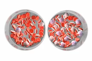
Normal and leukaemic blood, artwork C016 / 6261
Normal and leukaemic blood. Computer artwork comparing the appearance of normal blood (left) and that affected by leukaemia (right)

Background imageDiseased Collection: Effect of oxygen supply on tumour growth C016 / 6256

Effect of oxygen supply on tumour growth C016 / 6256
Effect of oxygen supply on tumour growth. Top: a small blood supply (tubes) means a low level of oxygen is available and the tumour (round) grows slowly

Background imageDiseased Collection: Cirrhosis of liver, light micrograph C016 / 0530

Cirrhosis of liver, light micrograph C016 / 0530
Cirrhosis of liver. Light micrograph of a section through liver tissue, damaged by cirrhosis. Cirrhosis occurs as a result of a range of factors causing damage to liver function

Background imageDiseased Collection: Bowel cancer, light micrograph C016 / 0511

Bowel cancer, light micrograph C016 / 0511
Bowel cancer. Light micrograph of a section through a carcinoid tumour in the small bowel. The tumour has invaded the submucosa of the bowel

Background imageDiseased Collection: Cirrhosis of liver, light micrograph C016 / 0529

Cirrhosis of liver, light micrograph C016 / 0529
Cirrhosis of liver. Light micrograph of a section through liver tissue, damaged by cirrhosis. Cirrhosis occurs as a result of a range of factors causing damage to liver function

Background imageDiseased Collection: Gingivitis, SEM C014 / 1443

Gingivitis, SEM C014 / 1443
Gingivitis. Coloured scanning electron micrograph (SEM) of a large number of bacteria (round) in the gingiva (gums) of a human mouth

Background imageDiseased Collection: Gingivitis, SEM C014 / 1444

Gingivitis, SEM C014 / 1444
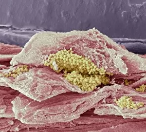
Gingivitis. Coloured scanning electron micrograph (SEM) of a large number of bacteria (yellow) in the gingiva (gums) of a human mouth

Background imageDiseased Collection: Mouth cancer cells, SEM

Mouth cancer cells, SEM
Mouth cancer cells. Coloured scanning electron micrograph (SEM) of squamous cell carcinoma (cancer) cells from a human mouth

Background imageDiseased Collection: Mouth cancer cell dividing, SEM

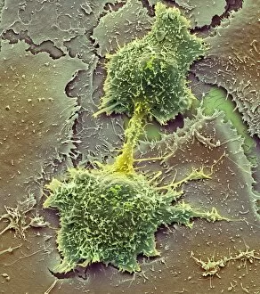
Mouth cancer cell dividing, SEM
Mouth cancer dividing. Coloured scanning electron micrograph (SEM) of a squamous cell carcinoma (cancer) cell from a human mouth undergoing mitosis (nuclear division)

Background imageDiseased Collection: Narrowed artery, artwork

Narrowed artery, artwork
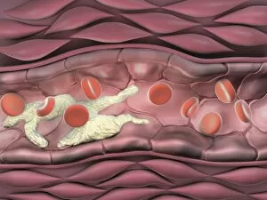
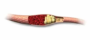
Narrowed artery. Computer artwork of a section through an artery (horizontal, centre) affected by atherosclerosis, showing red blood cells (red) and fat deposits (cream)

Background imageDiseased Collection: Mouth cancer cell, SEM

Mouth cancer cell, SEM
Mouth cancer cell. Coloured scanning electron micrograph (SEM) showing the surface of a squamous cell carcinoma (cancer) cell from a human mouth

Background imageDiseased Collection: Diseased cocoa pods, Ecuador C016 / 8421

Diseased cocoa pods, Ecuador C016 / 8421
Diseased cocoa pods. Cocoa (Theobroma cacao) fruit (pods) infected with frosty pod rot disease. This damaging disease is caused by the basidiomycete fungus Moniliophthora roreri

Background imageDiseased Collection: Secondary bowel cancer, endoscopic view C016 / 8337

Secondary bowel cancer, endoscopic view C016 / 8337
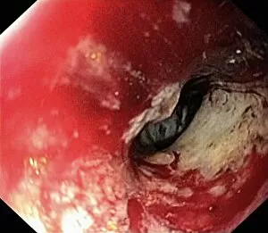
Secondary bowel cancer. Endoscopic view of the interior of a patients descending duodenum (first part of the small intestine), showing cancer that has spread (metastasised) from the pancreas

Background imageDiseased Collection: Intestinal lymphoid hyperplasia C016 / 8334

Intestinal lymphoid hyperplasia C016 / 8334
Intestinal lymphoid hyperplasia. Endoscopic view of lymphoid hyperplasia in the terminal ileum - the last part of the small intestine

Background imageDiseased Collection: Hiatal hernia, endoscopic view C016 / 8330

Hiatal hernia, endoscopic view C016 / 8330
Hiatal hernia. Endoscopic view of a hiatal hernia (gastro-oesophageal prolapse), seen from below, with the endoscope(black) seen in an inverted position inside the stomach

Background imageDiseased Collection: Hiatal hernia, endoscopic view C016 / 8329

Hiatal hernia, endoscopic view C016 / 8329
Hiatal hernia. Endoscopic view of a hiatal hernia (gastro-oesophageal prolapse), seen from above. A hernia is the protrusion of an organ, or the fascia of an organ

Background imageDiseased Collection: Hyperplastic polyps in the stomach C016 / 8327

Hyperplastic polyps in the stomach C016 / 8327
Hyperplastic polyps in the stomach. Endoscopic view of small hyperplastic polyps (growths) in the gastric fundus of the stomach

Background imageDiseased Collection: Diverticulum of the duodenum C016 / 8320

Diverticulum of the duodenum C016 / 8320
Diverticulum of the duodenum. Endoscope view of a diverticulum in the wall of the descending part of the duodenum - the first section of the small intestine

Background imageDiseased Collection: Crohns disease, endoscopic view

Crohns disease, endoscopic view
Crohns disease. Endoscopic view of the interior of a patients neoterminal ileum affected by Crohns disease, showing ulceration (white)

Background imageDiseased Collection: Ulcerative colitis, endoscopic view C016 / 8313

Ulcerative colitis, endoscopic view C016 / 8313
Ulcerative colitis. Endoscopic view of the interior of a patients distal colon (large intestine) affected by ulcerative colitis (UC), showing ulceration (white areas)

Background imageDiseased Collection: Antrum occult mucosal bleeding C016 / 8314

Antrum occult mucosal bleeding C016 / 8314
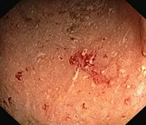
Antrum occult mucosal bleeding. Endoscopic view of the interior of a patients antrum (final portion of stomach), showing bleeding (red) of the mucosal lining

Background imageDiseased Collection: Ulcerative colitis, endoscopic view C016 / 8312

Ulcerative colitis, endoscopic view C016 / 8312
Ulcerative colitis. Endoscopic view of the interior of a patients distal colon (large intestine) affected by ulcerative colitis (UC), showing ulceration (white areas)

Background imageDiseased Collection: Ulcerative proctitis, endoscopic view C016 / 8311

Ulcerative proctitis, endoscopic view C016 / 8311
Ulcerative colitis. Endoscopic view of the interior of a patients anus, showing ulceration (white areas) cause by ulcerative proctitis

Background imageDiseased Collection: Ulcerative colitis, endoscopic view C016 / 8310

Ulcerative colitis, endoscopic view C016 / 8310
Ulcerative colitis. Endoscopic view of the interior of a patients colon (large intestine) affected by ulcerative colitis (UC), showing ulceration (white areas)

Background imageDiseased Collection: Ulcerative colitis, endoscopic view C016 / 8309

Ulcerative colitis, endoscopic view C016 / 8309
Ulcerative colitis. Endoscopic view of the interior of a patients colon (large intestine) affected by ulcerative colitis (UC), showing ulceration (white areas)

Background imageDiseased Collection: Ulcerative colitis, endoscopic view C016 / 8306

Ulcerative colitis, endoscopic view C016 / 8306
Ulcerative colitis. Endoscopic view of the interior of a patients distal colon (large intestine) affected by ulcerative colitis (UC), showing ulceration (white areas)

Background imageDiseased Collection: Colon cancer, endoscopic view C016 / 8307

Colon cancer, endoscopic view C016 / 8307
Colon cancer. Endoscopic view of a malignant (cancerous) adenocarcinoma (lower right) in a patients ascending colon (large intestine)

Background imageDiseased Collection: Melanosis coli, endoscopic view C016 / 8304

Melanosis coli, endoscopic view C016 / 8304
Melanosis coli. Endoscopic view of the interior of a patients colon affected by melanosis coli (pseudomelanosis coli). This benign condition causes brown pigmentation of the lining of the colon

Background imageDiseased Collection: Diseased organs, artwork

Diseased organs, artwork
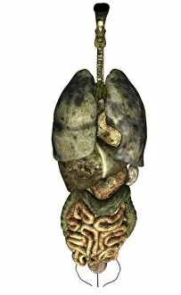
Diseased organs. Computer artwork showing diseased major organs of the human torso. From top to bottom: larynx (voicebox), trachea (windpipe), lungs (centre left and right), heart (centre), liver

Background imageDiseased Collection: Diabetes, conceptual image C013 / 5942

Diabetes, conceptual image C013 / 5942
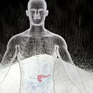
Diabetes, conceptual image. Computer artwork showing the location of the pancreas (pink) in the human body in front of a mound of sugar

Background imageDiseased Collection: Throat cancer, artwork C013 / 5939

Throat cancer, artwork C013 / 5939
Throat cancer. Cutaway computer artwork of a persons head, showing cancer (yellow) in the pharynx (centre) and oesophagus (lower centre)

Background imageDiseased Collection: Osteonecrosis of the jaw, artwork C017 / 8039

Osteonecrosis of the jaw, artwork C017 / 8039
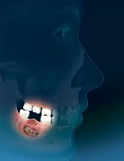
Osteonecrosis of the jaw. Computer artwork showing osteonecrosis (death of bone tissue) in a persons right mandible (lower jaw bone)

Background imageDiseased Collection: Atheromatous artery, artwork C015 / 6445

Atheromatous artery, artwork C015 / 6445
Atheromatous artery. Computer artwork showing the interior of an artery affected by atherosclerosis. Atherosclerosis is narrowing of an artery (stenosis) due to fatty deposits (beige)

Background imageDiseased Collection: Atheromatous artery, artwork C015 / 6446

Atheromatous artery, artwork C015 / 6446
Atheromatous artery. Cutaway computer artwork showing the interior of an artery affected by atherosclerosis. Atherosclerosis is narrowing of an artery (stenosis) due to fatty deposits (beige)

Background imageDiseased Collection: Single-kidney anatomy, artwork C016 / 2902

Single-kidney anatomy, artwork C016 / 2902
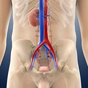
Single-kidney anatomy. Artwork showing an anterior (frontal) view of human kidneys in relation to the main torso blood vessels (vena cava, blue; aorta, red) and the male abdomen

Background imageDiseased Collection: Single-kidney anatomy, artwork C016 / 2903

Single-kidney anatomy, artwork C016 / 2903
Single-kidney anatomy. Artwork showing a posterior (from behind) view of human kidneys in relation to the main torso blood vessels (vena cava, blue; aorta, red) and the male abdomen

Background imageDiseased Collection: Single-kidney anatomy, artwork C016 / 2901

Single-kidney anatomy, artwork C016 / 2901
Single-kidney anatomy. Artwork showing an anterior (frontal) view of human kidneys in relation to the main torso blood vessels (vena cava, blue; aorta, red) and bones of the male skeleton

Background imageDiseased Collection: Single-kidney anatomy, artwork C016 / 2898

Single-kidney anatomy, artwork C016 / 2898
Single-kidney anatomy. Artwork showing an oblique right-frontal view of human kidneys in relation to the main torso blood vessels (vena cava, blue; aorta)

Background imageDiseased Collection: Single-kidney anatomy, artwork C016 / 2897

Single-kidney anatomy, artwork C016 / 2897
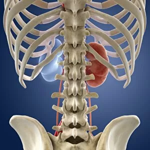
Single-kidney anatomy. Artwork showing a posterior (from behind) view of human kidneys in relation to the bones of the skeleton. One kidney (brown, upper right) has a normal appearance

Background imageDiseased Collection: Single-kidney anatomy, artwork C016 / 2900

Single-kidney anatomy, artwork C016 / 2900
Single-kidney anatomy. Artwork showing an anterior (frontal) view of human kidneys in relation to the main torso blood vessels (vena cava, blue; aorta, red) and bones of the skeleton

Background imageDiseased Collection: Single-kidney anatomy, artwork C016 / 2896

Single-kidney anatomy, artwork C016 / 2896
Single-kidney anatomy. Artwork showing an anterior (frontal) view of human kidneys in relation to the main torso blood vessels (vena cava, blue; aorta, red)


All Professionally Made to Order for Quick Shipping

"Diseased: A Glimpse into the World of Health Challenges" Bunions and X-ray: Unveiling the Painful Reality of Foot Deformities. Coral Spot Fungus on Sycamore Twig: Nature's Intricate Battle with Disease. Tension Pneumothorax Revealed: When Every Breath Becomes a Struggle. Bacterial Meningitis Unmasked: Peering into the Depths of an Invisible Threat. The Diseased Heart: An Organ in Need of Healing and Care. Malaria Parasites Under TEM Microscope: Understanding the Silent Invaders Within Us. Historic Advertisement for Migranin: Tracing Headache Relief Through Time and Culture. Consultation Hour at Sebastianeum Spa Hotel, Bavaria, 1880s: Seeking Remedies Amidst Serenity. The Spa Doctor in Thuringia, Germany: Bridging Medicine and Wellness in Centuries Past. At the Sickbed - A Mother's Vigilance Over Her Ill Child, France 1881: Love Transcends Disease's Grip. Bei der Dorfsybille - Prophetess from Germany 1881: Ancient Wisdom Meets Modern Maladies Caring for Casualties at Berlin Ambulance Station, 1895 Germany .