Fraud Blocker Skip to main content

Endocrinology Collection (page 3)

"Exploring the Intricacies of Endocrinology: From Goitre to Hormones" In the realm of medical science

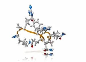
Background imageEndocrinology Collection: Obestatin molecule C014 / 4909

Obestatin molecule C014 / 4909
Obestatin molecule. Computer artwork showing the structure of a molecule of obestatin. Obestatin is thought to supress hunger and reduce food intake, thereby reducing weight gain

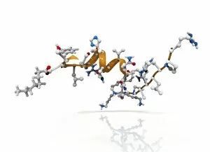
Background imageEndocrinology Collection: Activated ghrelin hormone molecule C014 / 4901

Activated ghrelin hormone molecule C014 / 4901
Activated ghrelin hormone molecule. Computer model showing the crystal structure of the human hormone ghrelin. The crystal structure consists of both the secondary structure

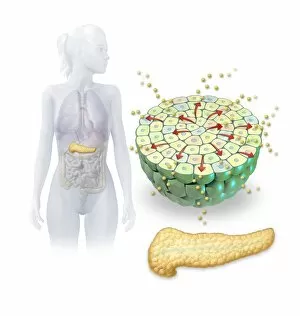
Background imageEndocrinology Collection: Insulin production, artwork

Insulin production, artwork
Insulin production. Computer artwork of the human pancreas (yellow) and an islet of Langerhans (green) producing the hormone insulin (small spheres)

Background imageEndocrinology Collection: Follicle-stimulating hormone complex C015 / 0945

Follicle-stimulating hormone complex C015 / 0945
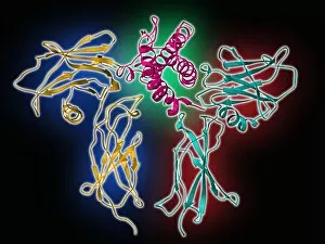
Follicle-stimulating hormone (FSH) complex with receptor, molecular model. FSH helps to regulate human sexual development and reproductive processes. In females, it acts on follicles in the ovaries

Background imageEndocrinology Collection: Follicle-stimulating hormone complex C015 / 0944

Follicle-stimulating hormone complex C015 / 0944
Follicle-stimulating hormone (FSH) complex with receptor, molecular model. FSH helps to regulate human sexual development and reproductive processes. In females, it acts on follicles in the ovaries

Background imageEndocrinology Collection: Melatonin, light micrograph

Melatonin, light micrograph
Melatonin. Light micrograph in polarised light of crystals of the hormone melatonin. Melatonin is secreted by the pineal gland in the brain that controls the bodys biological rhythm

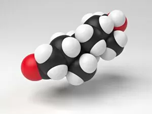
Background imageEndocrinology Collection: Testosterone hormone molecule

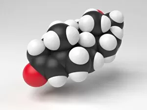
Testosterone hormone molecule. Computer model showing the structure of a molecule of the male sex hormone testosterone. Testosterone is the main human androgen

Background imageEndocrinology Collection: Progesterone hormone molecule

Progesterone hormone molecule. Computer model showing the structure of a molecule of the hormone progesterone. Progesterone is produced in the ovaries of women and the testes of men

Background imageEndocrinology Collection: Cortisol crystals, light micrograph C015 / 6786

Cortisol crystals, light micrograph C015 / 6786
Cortisol crystals, polarised light micrograph. Cortisol is a steroid hormone produced by the adrenal glands, which sit on top of the kidneys

Background imageEndocrinology Collection: Androgen receptor, molecular model C015 / 6113

Androgen receptor, molecular model C015 / 6113
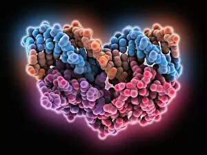
Androgen receptor. Molecular model of the DNA-binding region of an androgen receptor (purple and red) complexed with DNA (deoxyribonucleic acid, blue and orange)

Background imageEndocrinology Collection: Androgen receptor, molecular model C015 / 6112

Androgen receptor, molecular model C015 / 6112
Androgen receptor. Molecular model of the DNA-binding region of an androgen receptor (purple and brown) complexed with DNA (deoxyribonucleic acid, turquoise and red)

Background imageEndocrinology Collection: Urinary system, artwork C015 / 6455

Urinary system, artwork C015 / 6455
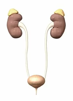
Urinary system. Computer artwork showing the organs of the human urinary system. The kidneys (red, top left and right) filter waste products such as urea from the blood (red and blue) to form urine

Background imageEndocrinology Collection: Urinary system, artwork C015 / 6454

Urinary system, artwork C015 / 6454
Urinary system. Computer artwork showing the organs of the human urinary system. The kidneys (red, top left and right) filter waste products such as urea from the blood to form urine

Background imageEndocrinology Collection: Thyroid gland, SEM C015 / 6419

Thyroid gland, SEM C015 / 6419
Thyroid gland. Coloured scanning electron micrograph (SEM) of a fracture through the thyroid gland revealing several follicles (green and blue). Between the follicles is connective tissue (pink)

Background imageEndocrinology Collection: Erythropoietin hormone complex C016 / 4461

Erythropoietin hormone complex C016 / 4461
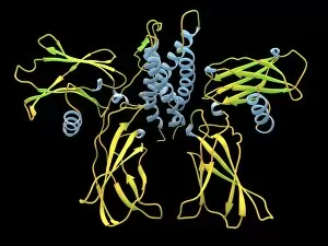
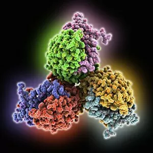
Erythropoietin hormone complex. Computer model showing the secondary structure of a molecule of the human hormone Erythropoietin (EPO), complexed with an erythropoetin receptor molecule

Background imageEndocrinology Collection: Erythropoietin hormone complex C016 / 4458

Erythropoietin hormone complex C016 / 4458
Erythropoietin hormone complex. Computer model showing the secondary structure of a molecule of the human hormone Erythropoietin (EPO), complexed with an erythropoetin receptor molecule

Background imageEndocrinology Collection: Erythropoietin hormone complex C016 / 4451

Erythropoietin hormone complex C016 / 4451
Erythropoietin hormone complex. Computer model showing the secondary structure of a molecule of the human hormone Erythropoietin (EPO), complexed with an erythropoetin receptor molecule

Background imageEndocrinology Collection: Follicle-stimulating hormone complex C016 / 2794

Follicle-stimulating hormone complex C016 / 2794
Follicle-stimulating hormone (FSH) complex with receptor, molecular model. FSH helps to regulate human sexual development and reproductive processes. In females, it acts on follicles in the ovaries

Background imageEndocrinology Collection: Follicle-stimulating hormone complex C016 / 2793

Follicle-stimulating hormone complex C016 / 2793
Follicle-stimulating hormone (FSH) complex with receptor, molecular model. FSH helps to regulate human sexual development and reproductive processes. In females, it acts on follicles in the ovaries

Background imageEndocrinology Collection: Human growth hormone molecule C013 / 9006

Human growth hormone molecule C013 / 9006
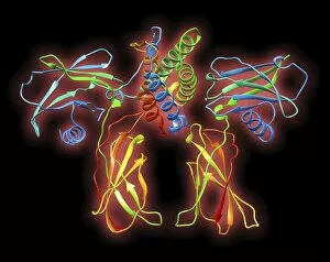
Human growth hormone. Molecular model of human growth hormone (hGH, yellow) bound to the extracellular domain of the human growth hormone binding protein (hGHBP)

Background imageEndocrinology Collection: Erythropoietin hormone complex

Erythropoietin hormone complex. Computer model showing the secondary structure of a molecule of the human hormone Erythropoietin (EPO), complexed with an erythropoetin receptor molecule

Background imageEndocrinology Collection: Kidneys and ureters, artwork C013 / 4672

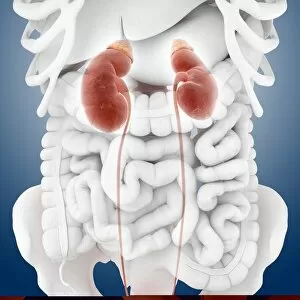
Kidneys and ureters, artwork C013 / 4672
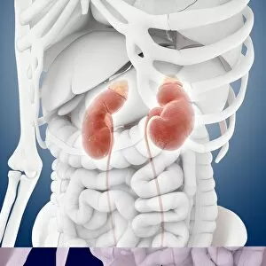
Kidneys and ureters. Computer artwork showing an oblique anterior view of the kidneys, the main organs of the urinary system, relative to the skeleton and abdominal organs (white)

Background imageEndocrinology Collection: Thyroid anatomy, artwork C013 / 4674

Thyroid anatomy, artwork C013 / 4674
Thyroid anatomy. Computer artwork of the thyroid glands (pink) of the neck, shown in relation to the bodys bones and throat structures (white)

Background imageEndocrinology Collection: Kidneys and ureters, artwork C013 / 4671

Kidneys and ureters, artwork C013 / 4671
Kidneys and ureters. Computer artwork showing an anterior view of the kidneys, the main organs of the urinary system, relative to the skeleton and abdominal organs (white)

Background imageEndocrinology Collection: Urinary system, artwork C013 / 4673

Urinary system, artwork C013 / 4673
Urinary system. Computer artwork showing an anterior view of the kidneys, the main organs of the urinary system, relative to the skeleton and abdominal organs (white)

Background imageEndocrinology Collection: Urinary system, artwork C013 / 4670

Urinary system, artwork C013 / 4670
Urinary system. Computer artwork showing the kidneys (top), the main organs of the urinary system. The kidneys are located in the upper abdomen

Background imageEndocrinology Collection: Urinary system, artwork C013 / 4669

Urinary system, artwork C013 / 4669
Urinary system. Computer artwork showing the kidneys (top), the main organs of the urinary system. The kidneys are located in the upper abdomen

Background imageEndocrinology Collection: Kidneys and ureters, artwork C013 / 4668

Kidneys and ureters, artwork C013 / 4668
Kidneys and ureters. Computer artwork showing the kidneys, the main organs of the urinary system. The kidneys are located in the upper abdomen

Background imageEndocrinology Collection: Thyroid gland, light micrograph

Thyroid gland, light micrograph
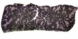
Thyroid gland. Coloured light micrograph of a thyroid gland showing the follicles. The follicles are lined by a single layer of cuboidal epithelial cells (purple, dark nuclei)

Background imageEndocrinology Collection: Thyroid gland follicle, TEM

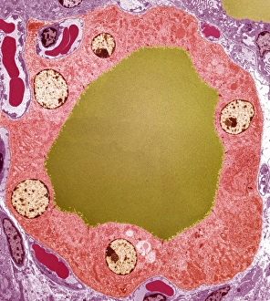
Thyroid gland follicle, TEM
Thyroid gland follicle. Coloured transmission electron micrograph (TEM) of a follicle from the thyroid gland. The follicle consists of a layer of epithelial cells (pink)

Background imageEndocrinology Collection: Endocrine cell, TEM

Endocrine cell, TEM
Endocrine cell. Coloured transmission electron micrograph (TEM) of a cultured endocrine cell. Endocrine cells are part of the endocrine system

Background imageEndocrinology Collection: Pituitary gland tissue, light micrograph

Pituitary gland tissue, light micrograph
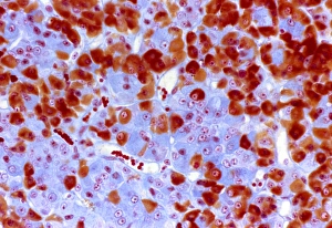
Pituitary gland tissue. Light micrograph of a section through the pars distalis, the anterior part of the adenohypophysis in the pituitary gland

Background imageEndocrinology Collection: Pituitary gland, TEM

Pituitary gland, TEM
Pituitary gland. Coloured transmission electron micrograph (TEM) of cells in the anterior pituitary gland, a hormone-secreting gland at the base of the brain

Background imageEndocrinology Collection: Kidneys and adrenal glands

Kidneys and adrenal glands
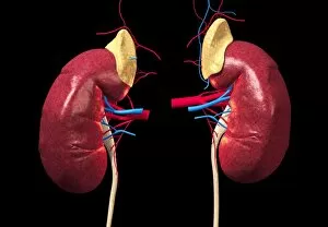
Kidneys. Computer artwork of the human renal system. The kidneys (brown/red, left and right) filter waste products and water from blood that passes through the organs in the renal arteries (red)

Background imageEndocrinology Collection: Pancreas

Pancreas. Computer artwork of a human pancreas. The pancreas is between 15 and 20 centimetres in length. It is located in the abdominal cavity behind the stomach

Background imageEndocrinology Collection: Thyroid follicles, artwork

Thyroid follicles, artwork
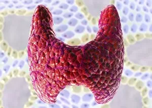
Thyroid gland. Computer artwork of a thyroid gland (red, centre), superimposed on an image of cells of thyroid follicles (green, blue)

Background imageEndocrinology Collection: Diabetes mellitus, computer artwork

Diabetes mellitus, computer artwork. Diabetes is caused by low levels of the hormone insulin and resistance to insulin by the body cells

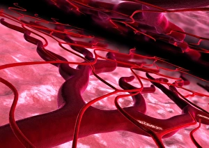
Background imageEndocrinology Collection: Blood vessel dilation, computer artwork

Blood vessel dilation, computer artwork
Blood vessel dilation. Computer artwork of blood vessel dilation (expansion) in response to drugs taken to control arterial blood pressure

Background imageEndocrinology Collection: Skin pigmentation in Addisons disease

Skin pigmentation in Addisons disease
Skin pigmentation from Addisons disease. Light micrograph of a section through the skin of a patient with Addisons disease
Background imageEndocrinology Collection: Charles Brown-Sequard, physiologist

Charles Brown-Sequard, physiologist

Background imageEndocrinology Collection: Thyroid gland, artwork

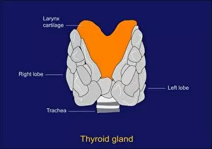
Thyroid gland, artwork. This two-lobed endocrine gland is found in the neck, in front of the trachea (windpipe, striped). The larynx (voice box, orange) sits above it

Background imageEndocrinology Collection: Thymus gland tissue, light micrograph

Thymus gland tissue, light micrograph
Thymus gland tissue. Light micrograph of a transverse section through tissue from the thymus gland, part of the lymphoid system

Background imageEndocrinology Collection: Islets of Langerhans cells, artwork

Islets of Langerhans cells, artwork
Islets of Langerhans cells, computer artwork. These cells (large, pink) of the pancreas release hormones (dots) into the blood
Background imageEndocrinology Collection: Hypothalamus and hypophysis, diagram

Hypothalamus and hypophysis, diagram
Hypothalamus and hypophysis. Diagram showing the brains hypothalamus (white) and hypophysis (pituitary gland, orange), the link between the nervous system and the endocrine system

Background imageEndocrinology Collection: Parathyroid glands, artwork

Parathyroid glands, artwork
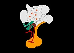
Artwork of the four small parathyroid glands lying on the thyroid glands in the neck of a male figure. The parathyroid glands (light pink) produce calcitonin

Background imageEndocrinology Collection: Hypothalamus and hypophysis, artwork

Hypothalamus and hypophysis, artwork
Hypothalamus and hypophysis. Artwork showing the brains hypothalamus (white) and hypophysis (pituitary gland, orange), the link between the nervous system and the endocrine system

Background imageEndocrinology Collection: Melatonin brain function, artwork

Melatonin brain function, artwork
Melatonin brain function. Artwork showing how the hormone melatonin (blue spheres) is secreted in the brain and helps to control the bodys biological clock

Background imageEndocrinology Collection: Synaptic endorphins, artwork

Synaptic endorphins, artwork
Synaptic endorphins. Artwork showing the release (top) of neurotransmitters called endorphins (yellow) from the end of a nerve (blue)


All Professionally Made to Order for Quick Shipping

"Exploring the Intricacies of Endocrinology: From Goitre to Hormones" In the realm of medical science, endocrinology stands as a fascinating field that unravels the secrets behind our body's intricate hormonal system. Journeying back in time, we encounter a 15th-century artwork depicting a haunting goitre, shedding light on historical thyroid disorders. Delving deeper into this captivating subject, we explore cortisol crystals through a mesmerizing light micrograph. These tiny formations offer us a glimpse into the inner workings of our stress response and adrenal function. Moving forward, we stumble upon another intriguing light micrograph showcasing the delicate Islet of Langerhans. Nestled within our pancreas, these clusters of cells play an essential role in regulating blood sugar levels and are vital for managing diabetes. Shifting gears to neurotransmitters, we encounter oxytocin - often referred to as the "love hormone. " This molecule holds immense power over human bonding and social interactions, highlighting its significance in understanding emotional connections. Our exploration takes us further into thyroid anatomy with yet another captivating artwork. The intricate details depicted guide us through this crucial gland's structure and its impact on metabolism regulation. Returning to Islets of Langerhans but from an artistic perspective this time; their vibrant portrayal captures their importance in maintaining glucose balance within our bodies—a testament to both scientific beauty and functionality. Once again encountering cortisol crystals under microscopic lenses reveals more about stress-related conditions while reminding us how interconnected hormones are with our overall well-being. A poignant engraving portrays a woman suffering from congenital iodine deficiency syndrome or cretinism due to inadequate iodine intake during pregnancy—an alarming reminder of how nutrition plays an integral role in endocrine health. Crossing paths with an anatomical cross-section image showcases the complexity of adrenal glands—tiny powerhouses responsible for producing various hormones that regulate numerous bodily functions such as metabolism and stress response.