Fraud Blocker Skip to main content

Infection Collection (page 10)

"Infection: A Historical Journey into the Battle against Disease" In the realm of medicine, it has long been a formidable adversary

Background imageInfection Collection: History of medicine. Dentist. Middle Ages. Satire. Engraving
History of medicine. Dentist. Middle Ages. Satire. Engraving
History of medicine. Dentist (barbers or general physicians). Middle Ages. Satire. Engraving of a collection of comic work. 12th century. Colored

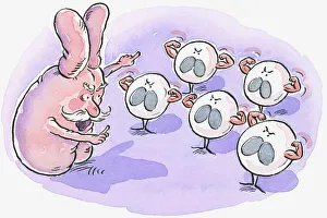
Background imageInfection Collection: Cartoon representing thymus pointing at white blood cells flexing muscles

Cartoon representing thymus pointing at white blood cells flexing muscles

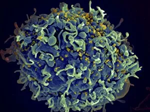
Background imageInfection Collection: Scanning electron micrograph of HIV particles infecting a human H9 T cell

Scanning electron micrograph of HIV particles infecting a human H9 T cell

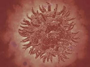
Background imageInfection Collection: Stylized rabies virus particles

Stylized rabies virus particles, the cause of the viral neuroinvasive disease acute encephalitis

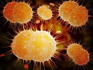
Background imageInfection Collection: Microscopic view of Neisseria gonorrhoeae

Microscopic view of Neisseria gonorrhoeae. Neisseria gonorrhoeae typically infects the mucous membranes causing infections such as urethritis, cervicitis, salpingitis, pelvic inflammatory disease

Background imageInfection Collection: Microscopic view of yellow fever virus

Microscopic view of yellow fever virus. Yellow fever is an acute viral disease

Background imageInfection Collection: Microscopic view of Giardiasis

Microscopic view of Giardiasis, an infectious disease caused by a unicellular parasite known as Giardia lamblia

Background imageInfection Collection: Microscopic view of bacterial pneumonia

Microscopic view of bacterial pneumonia. Bacterial pneumonia is a type of pneumonia caused by bacterial infection. Pneumonia can be generally defined as inflammation of the lung parenchyma

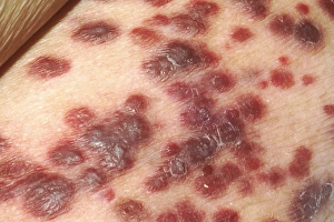
Background imageInfection Collection: Kaposis sarcoma on the skin of an AIDS patient

Kaposis sarcoma on the skin of an AIDS patient

Background imageInfection Collection: Microscopic view of Streptococcus pneumoniae

Microscopic view of Streptococcus pneumoniae, also known as pneumococcus. Pneumococcus is a Gram-positive coccus shaped pathogenic bacteria which causes many types of pneumococcal infections in

Background imageInfection Collection: Anatomy of foot fungus with microscopic close-up

Anatomy of foot fungus with microscopic close-up

Background imageInfection Collection: Conceptual image of rabies virus

Conceptual image of rabies virus

Background imageInfection Collection: Can t Pound It Into His Head! American cartoon by Dr. Seuss (Theodor Geisel) for PM

Can t Pound It Into His Head! American cartoon by Dr. Seuss (Theodor Geisel) for PM, 30 September 1942
CARTOON: WORLD WAR II. Can t Pound It Into His Head! American cartoon by Dr. Seuss (Theodor Geisel) for PM, 30 September 1942

Background imageInfection Collection: HIV infected macrophage, SEM C018 / 8598

HIV infected macrophage, SEM C018 / 8598
HIV infected macrophage. Coloured ion-abrasion scanning electron micrograph (IA-SEM) of a macrophage white blood cell infected with human immunodeficiency virus (HIV, red)

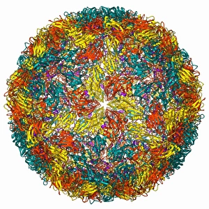
Background imageInfection Collection: Foot-and-mouth disease virus F006 / 9556

Foot-and-mouth disease virus F006 / 9556
Foot-and-mouth disease virus. Molecular model of the foot-and-mouth disease (FMD) virus (Aphtae epizooticae) protein coat (capsid)

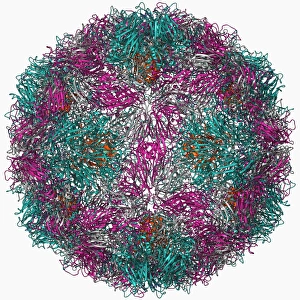
Background imageInfection Collection: Rhinovirus 16 capsid, molecular model F006 / 9431

Rhinovirus 16 capsid, molecular model F006 / 9431
Rhinovirus 16 capsid, molecular model. This is human rhinovirus 16. The rhinovirus infects the upper respiratory tract and is the cause of the common cold. It is spread by coughs and sneezes

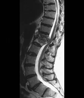
Background imageInfection Collection: Tuberculosis of the spine, MRI scan

Tuberculosis of the spine, MRI scan
Tuberculosis of the spine. Magnetic resonance imaging (MRI) scan of a sagittal section through the spine of a 74-year-old patient with tuberculosis (TB) of the spine, or Potts disease

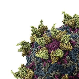
Background imageInfection Collection: Rhinovirus and antibody, molecular model C015 / 7138

Rhinovirus and antibody, molecular model C015 / 7138
Rhinovirus. Molecular model of the antigen-binding fragment (Fab) from a strongly neutralising antibody bound to a human rhinovirus 14 (HRV-14) particle

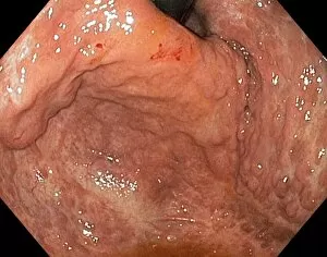
Background imageInfection Collection: Severe gastritis, endoscope view C016 / 3898

Severe gastritis, endoscope view C016 / 3898
Severe gastritis, endoscope view. Gastritis is inflammation of the stomach lining. It causes pain on eating or drinking, and may cause nausea and vomiting

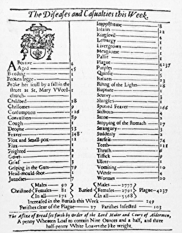
Background imageInfection Collection: A Bill of Mortality for the London plague, week of 15 to 22 August 1665

A Bill of Mortality for the London plague, week of 15 to 22 August 1665. Of the 5, 568 deaths recorded, 4
MORTALITY BILL, 1665. A Bill of Mortality for the London plague, week of 15 to 22 August 1665. Of the 5, 568 deaths recorded, 4, 237 were attributed to the plague

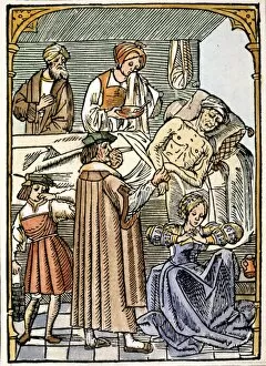
Background imageInfection Collection: A physician visits a plague victim, holding a sponge soaked in aromatic vinegar to his nose while

A physician visits a plague victim, holding a sponge soaked in aromatic vinegar to his nose while his assistant holds
PHYSICIAN & PLAGUE VICTIM. A physician visits a plague victim, holding a sponge soaked in aromatic vinegar to his nose while his assistant holds his own nose shut with his fingers

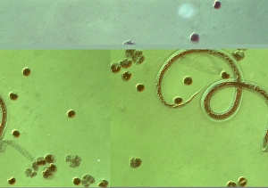
Background imageInfection Collection: Wuchereria bancrofti parasite

Wuchereria bancrofti parasite
Wuchereria bancrofti. Light micrograph of the microfilaria larval stage of the parasitic worm Wuchereria bancrofti, which causes filariasis in humans. W

Background imageInfection Collection: Eyelash hairs, SEM

Eyelash hairs, SEM
Eyelash hairs and skin. Coloured scanning electron micrograph (SEM) of eyelash hairs growing from the surface of human skin

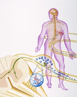
Background imageInfection Collection: Shingles nerve damage

Shingles nerve damage

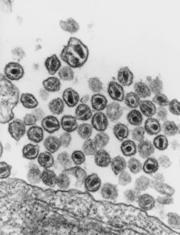
Background imageInfection Collection: TEM of HIV (AIDS) viruses budding from a T-cell

TEM of HIV (AIDS) viruses budding from a T-cell
AIDS virus. Transmission electron micrograph (TEM) of a T-lymphocyte blood cell infected with Human Immunodeficiency virus (HIV), the causative agent of AIDS

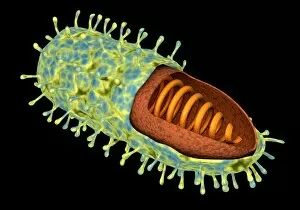
Background imageInfection Collection: AIDS virus

AIDS virus. Conceptual computer artwork of many HIV (human immunodeficiency virus) particles that are sectioned to show their internal structures

Background imageInfection Collection: Cholera prevention, satirical artwork

Cholera prevention, satirical artwork
Cholera prevention. Satirical artwork portraying a womans efforts to protect herself during an outbreak of cholera. Cholera is a bacterial infection of the intestines that was often fatal before

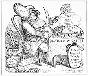
Background imageInfection Collection: Cholera doctor, satirical artwork

Cholera doctor, satirical artwork
Cholera doctor. Satirical artwork of a London doctor gaining wealth (money at lower left) during an 1835 cholera epidemic

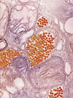
Background imageInfection Collection: Eastern equine encephalitis virus, TEM

Eastern equine encephalitis virus, TEM
Eastern equine encephalitis virus. Coloured transmission electron micrograph (TEM) of eastern equine encephalitis (EEE) virus particles (Alphavirus sp. orange) in the salivary gland of a mosquito

Background imageInfection Collection: HIV particles, TEM

HIV particles, TEM
HIV (human immunodeficiency virus) particles, coloured transmission electron micrograph (TEM). Each particle consists of an RNA (ribonucleic acid, purple/brown) genome

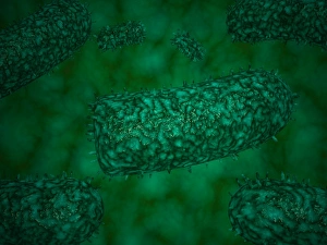
Background imageInfection Collection: Bacterial lung infection

Bacterial lung infection, computer artwork. Rod-shaped bacteria (bacilli, green) on the surface of the alveoli of the lungs (purple)

Background imageInfection Collection: Lymphocytes in hair follicle, SEM

Lymphocytes in hair follicle, SEM
Lymphocytes in hair follicle, coloured scanning electron micrograph (SEM). Large numbers of lymphocytes (yellow) are seen in inflamed and necrotic tissue at the base of hair shafts (red)

Background imageInfection Collection: Bacillus subtilis bacteria, SEM

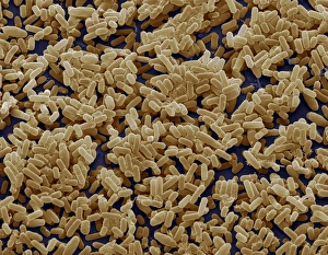
Bacillus subtilis bacteria, SEM
Bacillus subtilis. Coloured scanning electron micrograph (SEM) of Bacillus subtilis bacteria. B. subtilis, or hay bacillus, is an aerobic, gram-positive bacillus(rod-shaped bacterium)

Background imageInfection Collection: Bacteria on lung hairs

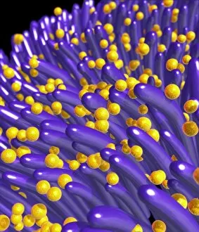
Bacteria on lung hairs
Bacteria in a lung. Computer artwork of bacteria (yellow) on cilia (hair-like structures) in a lung. Bacterial infection of the lungs can cause potentially fatal pneumonia

Background imageInfection Collection: White Oak School, Swanley, Kent

White Oak School, Swanley, Kent
General view of the White Oak School, Swanley, Kent. The school was established in 1903 by the Metropolitan Asylums Board for the treatment of ophthalmia, a highly infectious condition of the eyes

Background imageInfection Collection: Enfield Isolation Hospital, Winchmore Hill, Middlesex

Enfield Isolation Hospital, Winchmore Hill, Middlesex
The Isolation Hospital at Winchmore Hill, Enfield, Middlesex. The hospital, opened in 1900 by Enfield Urban District Council, later became known as South Lodge Hospital

Background imageInfection Collection: Princess Marys Hospital, Margate, Kent

Princess Marys Hospital, Margate, Kent
Entrance to the Princess Marys Hospital, Wilderness Road, Margate, Kent. The site, originally known as Eastcliff House, was acquired in 1898 by the Metropolitan Asylums Board as a seaside hospital

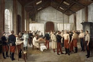
Background imageInfection Collection: Vaccination: c1895. Alfred Touchemolin (1829-1907) French painter. French army recruits

Vaccination: c1895. Alfred Touchemolin (1829-1907) French painter. French army recruits being vaccinated with Cowpox to protect them from the more virulent infection, Smallpox

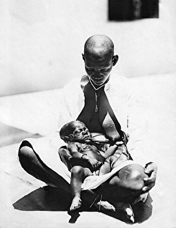
Background imageInfection Collection: Child with Yaws, Mombasa, Kenya, East Africa

Child with Yaws, Mombasa, Kenya, East Africa. 1923

Background imageInfection Collection: A-Tich-oo! Influenza in 1918

A-Tich-oo! Influenza in 1918
Influenza personified in the shape of a rather monstrous being tapping an unsuspecting chap on the head and announcing, Good evening! I m the new influenza. Date: 1918

Background imageInfection Collection: Advert for kruschen saits - a-tish-oo! 1918

Advert for kruschen saits - a-tish-oo! 1918
Don t be a afraid to sneeze ! Every grown-up, man and woman alike, should know the value of the Kruschen Habit as a safe, sure simple remedy against all inflammations of which the pus-germ-pace

Background imageInfection Collection: Advert for Evans Pastilles against influenza infection 1918

Advert for Evans Pastilles against influenza infection 1918
Take one in time Between January 1918 to December 1920 the influenza pandemic killed millions of mainly young healthy adults

Background imageInfection Collection: Advert for Sanitas - family antiseptic for wounds 1937

Advert for Sanitas - family antiseptic for wounds 1937
Just a small wound but.... I never run the risk of blood poisoning. As soon as any member of my family gets a scratch or cut, let alone a more serious wound, I instantly apply Sanitas

Background imageInfection Collection: SMALLPOX PATIENT. Smallpox patient in a hospital bed, early 20th century

SMALLPOX PATIENT. Smallpox patient in a hospital bed, early 20th century

Background imageInfection Collection: TUBERCULOSIS CAMP. Camp for children with tuberculosis

TUBERCULOSIS CAMP. Camp for children with tuberculosis. Photograph, American, early 20th century

Background imageInfection Collection: ILLINOIS: TUBERCULOSIS CAMP. Patient sitting outside a cabin at a tuberculosis camp in Illinois

ILLINOIS: TUBERCULOSIS CAMP. Patient sitting outside a cabin at a tuberculosis camp in Illinois, early 20th century

Background imageInfection Collection: ILLINOIS: TUBERCULOSIS CAMP. Patients outside a tent at a tuberculosis camp in Illinois

ILLINOIS: TUBERCULOSIS CAMP. Patients outside a tent at a tuberculosis camp in Illinois, early 20th century

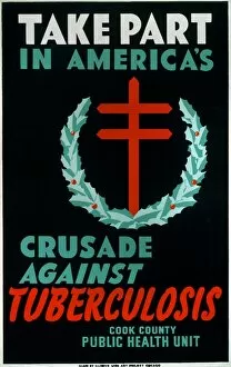
Background imageInfection Collection: NEW DEAL: WPA POSTER. Take Part In Americas Crusade Against Tuberculosis

NEW DEAL: WPA POSTER. Take Part In Americas Crusade Against Tuberculosis. City of Chicago Municipal Tuberculosis Sanitarium poster promoting tuberculosis testing


All Professionally Made to Order for Quick Shipping

"Infection: A Historical Journey into the Battle against Disease" In the realm of medicine, it has long been a formidable adversary. From ancient plagues to modern epidemics, humanity has faced countless battles against unseen foes that threaten our very existence. This caption takes us on a captivating journey through history, exploring various artworks and moments that shed light on the fight against infectious diseases. The Cow-Pock satirical etching by James Gillray in 1802 introduces us to Edward Jenner and his groundbreaking work on vaccination. This pivotal moment marked a turning point in medical history, as Jenner's discovery paved the way for immunization practices that would save countless lives. Moving further back in time, we encounter an eerie 17th-century artwork depicting a plague doctor. These haunting figures were tasked with treating victims during one of history's most devastating pandemics - the Black Death. Their iconic beaked masks and dark robes serve as chilling reminders of the horrors unleashed by infectious diseases. Fast forward to 1866, where Deaths Dispensary cartoon highlights water pollution as a source of disease. This powerful image serves as a stark reminder of how environmental factors can contribute to widespread infections and emphasizes the importance of clean water for public health. Another striking woodcut from London during the Great Plague of 1665 begs for divine intervention: "Lord, have mercy on London. " The desperation felt during this catastrophic event is palpable even centuries later, reminding us of our vulnerability when faced with rampant infections. Shifting gears to Cuba in c1900, we witness Dr. Carlos Finlay and Dr. Walter Reed leading efforts against yellow fever after the Spanish-American War. The oil painting captures their determination alongside other physicians observing inoculation procedures – showcasing bravery amidst uncertainty while combating deadly outbreaks. Calots spinal surgery illustration from the 19th century showcases medical advancements aimed at tackling specific infections affecting vital organs like tuberculosis or addressing conditions requiring surgical intervention such as spinal disorders.