
Inhibiting Collection
"Inhibiting: Unlocking the Secrets of Molecular Control" In the intricate world of molecular biology


All Professionally Made to Order for Quick Shipping
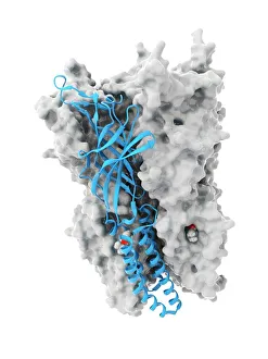
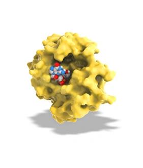
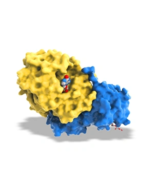
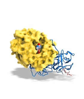
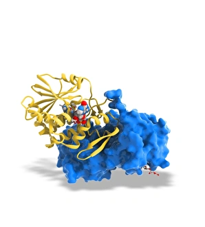
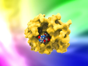
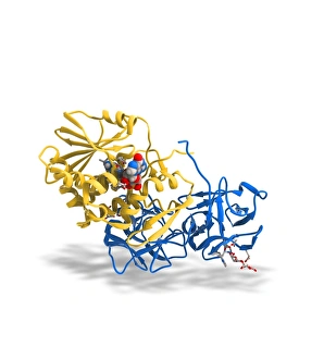
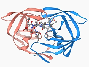
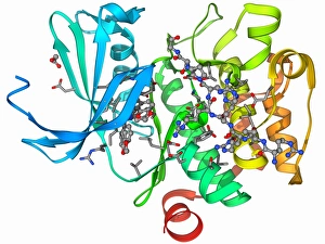
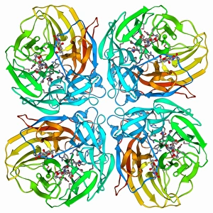
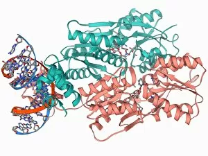
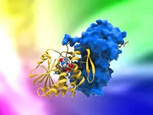
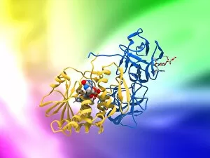
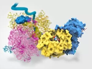
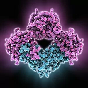
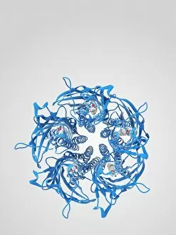
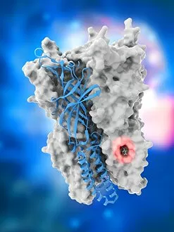
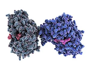
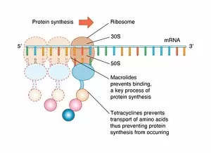
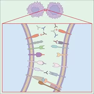
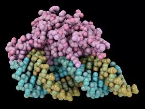
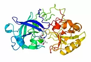
"Inhibiting: Unlocking the Secrets of Molecular Control" In the intricate world of molecular biology, inhibiting certain processes can hold the key to understanding and manipulating various biological functions. From anaesthetics blocking ion channels to potent inhibitors combating deadly viruses, scientists delve into these captivating mechanisms. Captured in stunning artwork, we witness an anaesthetic molecule skillfully inhibiting an ion channel (C015 / 6718). This interaction disrupts nerve signals, leading to temporary loss of sensation during medical procedures. The enigmatic ricin A-chain (C017 / 3653) takes center stage as it showcases its lethal potential. Its ability to inhibit protein synthesis within cells makes it a formidable toxin. The intricately designed ricin molecules (C017 / 3652-3651) further emphasize their dangerous nature. However, science also harnesses inhibition for positive purposes. HIV-1 protease and inhibitor complex (F006 / 9773) represents a breakthrough in antiretroviral therapy by targeting viral enzymes crucial for replication. Similarly, the kinase inhibitor complex (F006 / 9760) offers hope in cancer treatment by hindering abnormal cell growth pathways. As we explore further, we encounter a fascinating battle between a flu virus surface protein and a drug molecule (F006 / 9745). Inhibition prevents viral entry into host cells and potentially curbs influenza outbreaks. The HIV reverse transcription enzyme (F006 / 9684), responsible for converting viral RNA into DNA within human cells, faces resistance from inhibitors aiming to impede its activity. These inhibitors play a vital role in managing HIV infections worldwide. Lastly, we uncover the intriguing concept of RNA interference through the interplay between a viral suppressor and RNA molecules (F006/9488). By inhibiting gene expression at specific points along the genetic pathway, researchers unlock new possibilities for treating genetic disorders or combating harmful viruses.