Fraud Blocker Skip to main content

Microscope Collection (page 14)

"Unveiling the Unseen: Exploring the Microscopic World" Step into a realm of hidden wonders

Background imageMicroscope Collection: Inverted microscope

Inverted microscope
Medicine - Inverted microscope (Turin, University of Biology)

Background imageMicroscope Collection: Laser beam of stereo microscope device used in macular disease

Laser beam of stereo microscope device used in macular disease
Medicine - Laser beam of stereo microscope device used in macular disease

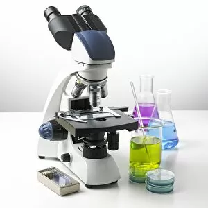
Background imageMicroscope Collection: Microscope, beaker, scalpel, tweezers, and chemistry samples on petri dish, close-up

Microscope, beaker, scalpel, tweezers, and chemistry samples on petri dish, close-up

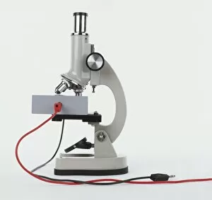
Background imageMicroscope Collection: Side view of a microscope with red and black wires

Side view of a microscope with red and black wires

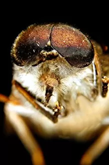
Background imageMicroscope Collection: Tabanidae. Horse Fly
Tabanidae. Horse Fly

Background imageMicroscope Collection: Sample of human bladder tissue seen through microscope, close-up

Sample of human bladder tissue seen through microscope, close-up

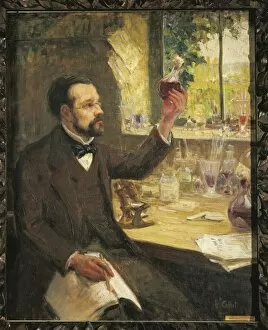
Background imageMicroscope Collection: France, Dole, Louis Pasteur (1822-1895) studying the fermentation of grape juice

France, Dole, Louis Pasteur (1822-1895) studying the fermentation of grape juice
France - 19th century. Louis Pasteur (1822-1895) studying the fermentation of grape juice. Portrait by Callot

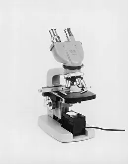
Background imageMicroscope Collection: Microscope

Microscope

Background imageMicroscope Collection: Using USB microscope to display magnified image of caterpillar on laptop screen

Using USB microscope to display magnified image of caterpillar on laptop screen

Background imageMicroscope Collection: Microscope Optics and Mirror Diagrams C017 / 3456

Microscope Optics and Mirror Diagrams C017 / 3456
Plate from 18th century encyclopedia showing illustrations of microscope optics and apparatus. Several technical innovations make microscopes better and easier to handle

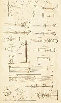
Background imageMicroscope Collection: Reflaction and Telescope Optics C017 / 3507

Reflaction and Telescope Optics C017 / 3507
Plate from 18th century encyclopedia showing diagrams of telescope optics, and light raypaths through prisms and lenses to the eye

Background imageMicroscope Collection: Food research, conceptual image

Food research, conceptual image

Background imageMicroscope Collection: Biological research F006 / 7189

Biological research F006 / 7189
Biological research

Background imageMicroscope Collection: Food research, conceptual image F006 / 7233

Food research, conceptual image F006 / 7233
Food research, conceptual image

Background imageMicroscope Collection: Food research, conceptual image F006 / 7232

Food research, conceptual image F006 / 7232
Food research, conceptual image

Background imageMicroscope Collection: Microbiology research, artwork F007 / 8429

Microbiology research, artwork F007 / 8429
Microbiology research, computer artwork

Background imageMicroscope Collection: Biological research, conceptual image F007 / 8172

Biological research, conceptual image F007 / 8172
Biological research, conceptual image

Background imageMicroscope Collection: Biological research F008 / 2162

Biological research F008 / 2162
Biological research

Background imageMicroscope Collection: Light microscope F008 / 2043

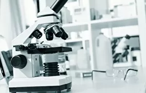
Light microscope F008 / 2043
Light microscope

Background imageMicroscope Collection: Biological research F008 / 3115

Biological research F008 / 3115
MODEL RELEASED. Biological research

Background imageMicroscope Collection: Microscope slide F008 / 2109

Microscope slide F008 / 2109
Microscope slide

Background imageMicroscope Collection: Microscope slide F008 / 2118

Microscope slide F008 / 2118
Microscope slide

Background imageMicroscope Collection: Biological research F008 / 2073

Biological research F008 / 2073
MODEL RELEASED. Biological research

Background imageMicroscope Collection: Microscopy sample F008 / 2110

Microscopy sample F008 / 2110
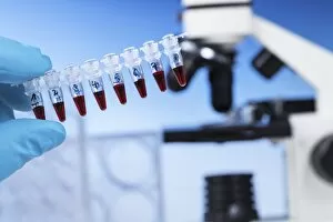
fluid sample in the test tube against a microscope

Background imageMicroscope Collection: Microscopy F008 / 2042

Microscopy F008 / 2042
Microscopy

Background imageMicroscope Collection: Blood smears F008 / 2107

Blood smears F008 / 2107
Blood smears on microscope slides

Background imageMicroscope Collection: Biological research F008 / 3116

Biological research F008 / 3116
MODEL RELEASED. Biological research

Background imageMicroscope Collection: Microscope slides F008 / 2108

Microscope slides F008 / 2108
Microscope slides

Background imageMicroscope Collection: Chemistry research F008 / 2052

Chemistry research F008 / 2052
Chemistry research

Background imageMicroscope Collection: Biological research F008 / 2044

Biological research F008 / 2044
MODEL RELEASED. Biological research

Background imageMicroscope Collection: Biological research F008 / 3113

Biological research F008 / 3113
MODEL RELEASED. Biological research

Background imageMicroscope Collection: Biological research F008 / 3114

Biological research F008 / 3114
MODEL RELEASED. Biological research

Background imageMicroscope Collection: Biological research F008 / 3112

Biological research F008 / 3112
MODEL RELEASED. Biological research

Background imageMicroscope Collection: IVF treatment F008 / 3572

IVF treatment F008 / 3572
IVF treatment. Embryo culture dish used for in vitro fertilisation (IVF)

Background imageMicroscope Collection: Optical light microscope with camera

Optical light microscope with camera
Microscope. Computer artwork of a typical optical microscope with a digital camera attached for photography. Below the camera these are the two eyepieces (binocular) for viewing the specimen

Background imageMicroscope Collection: Optical light microscope, artwork

Optical light microscope, artwork
Microscope. Computer artwork of a typical optical microscope. Below the camera these are the two eyepieces (binocular) for viewing the specimen

Background imageMicroscope Collection: Scanning tunnelling microscope, artwork F005 / 0813

Scanning tunnelling microscope, artwork F005 / 0813
Scanning tunnelling microscope (STM), computer artwork. The STM uses a fine needle with an electric potential running through it to scan the surface of an object

Background imageMicroscope Collection: Biological research F005 / 0936

Biological research F005 / 0936
MODEL RELEASED. Biological research. Researcher using a light microscope

Background imageMicroscope Collection: Biological research F005 / 0937

Biological research F005 / 0937
MODEL RELEASED. Biological research. Researcher using a light microscope

Background imageMicroscope Collection: H1N1 swine flu virus, TEM C018 / 8602

H1N1 swine flu virus, TEM C018 / 8602
H1N1 swine flu virus. Coloured transmission electron micrograph (TEM) of swine influenza (flu) virus particles (virions, yellow)

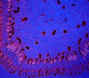
Background imageMicroscope Collection: Cotton stem, confocal micrograph C014 / 4636

Cotton stem, confocal micrograph C014 / 4636
Cotton stem. Laser scanning confocal micrograph (LSCM) of a section through the stem of a cotton (Gossypium sp.) plant

Background imageMicroscope Collection: Light microscope C014 / 1407

Light microscope C014 / 1407
Light microscope, seen in silhouetted profile. This microscope has a binocular eyepiece (an eyepiece for each eye). The sample is placed on a glass slide on the mount below centre

Background imageMicroscope Collection: Microscopic Vegetable Objects C017 / 3506

Microscopic Vegetable Objects C017 / 3506
Plate from 18th century encyclopedia showing illustrations of microscopical objects. Shown are various seeds of plants including; apple, clematis, columbine, cabbage, cucumber, jasmine, pear, poppy

Background imageMicroscope Collection: Biological research F007 / 0378

Biological research F007 / 0378
Biological research

Background imageMicroscope Collection: Light microscope F007 / 0356

Light microscope F007 / 0356
Light microscope

Background imageMicroscope Collection: Microbiology research F007 / 0304

Microbiology research F007 / 0304
MODEL RELEASED. Microbiology research

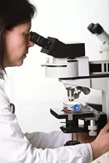
Background imageMicroscope Collection: Scientist using a light microscope F006 / 9023

Scientist using a light microscope F006 / 9023
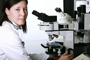
MODEL RELEASED. Scientist using a light microscope

Background imageMicroscope Collection: Biological research F006 / 9024

Biological research F006 / 9024
MODEL RELEASED. Biological research


All Professionally Made to Order for Quick Shipping

"Unveiling the Unseen: Exploring the Microscopic World" Step into a realm of hidden wonders, where scientists like Rosalind Franklin and Santiago Ramon y Cajal have unraveled the mysteries through their microscopic lens. From Liver Fluke to Norovirus particles, these pioneers have paved the way for groundbreaking discoveries. Witness the intricate beauty of Snail teeth and Fat cells as they come alive under the microscope's gaze. Marvel at Lubbock's Graphic 84, a visual masterpiece capturing the essence of this microcosmic universe. Delve deeper into this microscopic world and encounter Plasmodium sp. , a malarial parasite that has plagued humanity for centuries. Explore E. Coli bacterium in all its glory, revealing both its potential dangers and scientific significance. Through microscope slide preparation techniques, we gain insight into complex organisms like never before. Discover how Fleming used his photographic skills to document crucial moments in microbiology history. The microscope acts as our window into an unseen dimension - it allows us to explore realms beyond our naked eye's reach, and is through this powerful tool that we continue to unravel nature's secrets one magnified image at a time. So join us on this journey "Under the Microscope, " where every slide holds a story waiting to be told – stories that shape our understanding of life itself.