Fraud Blocker Skip to main content

Microscopic Collection (page 2)

"Exploring the Microscopic World: A Glimpse into Nature's Hidden Wonders" Step into a realm unseen by the naked eye

Background imageMicroscopic Collection: Microscopic Marine Life

Microscopic Marine Life
Various forms of microscopic marine life, described by Sibly as Animalcules Date: 1794

Background imageMicroscopic Collection: Scanning Electron Micrograph (SEM): Mosquito, Female; Magnification x 55 (A4 size: 29. 7 cm width)

Scanning Electron Micrograph (SEM): Mosquito, Female; Magnification x 55 (A4 size: 29. 7 cm width)
LRDS-83 Mosquito, Female Scanning Electron Micrograph (SEM) Anopheles sp. Magnification x 55 (A4 size: 29.7 cm width) Coloured by hand to enhance natural features

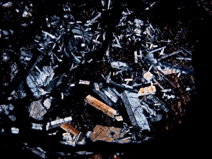
Background imageMicroscopic Collection: Microscope image of the Pasamonte eucrite

Microscope image of the Pasamonte eucrite
Microscopic image of the Pasamonte eucrite showing a basaltic texture. Field of view is 2.5mm across

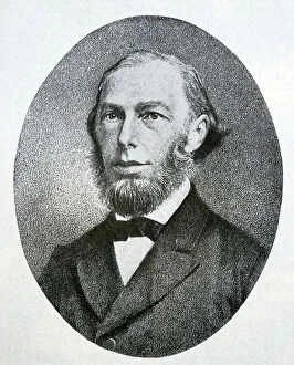
Background imageMicroscopic Collection: Max Johann Sigismund Schultze, 1850

Max Johann Sigismund Schultze, 1850
1576982 Max Johann Sigismund Schultze, 1850; (add.info.: Max Johann Sigismund Schultze (March 25, 1825 - January 16, 1874) was a German microscopic anatomist

Background imageMicroscopic Collection: Magnified image of common moss, Phasum cuspidatum 1, and microscopic enlargment of pond algae

Magnified image of common moss, Phasum cuspidatum 1, and microscopic enlargment of pond algae
FLO4574183 Magnified image of common moss, Phasum cuspidatum 1, and microscopic enlargment of pond algae. Handcoloured copperplate engraving from Bertuch's " Bilderbuch fur Kinder"

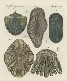
Background imageMicroscopic Collection: Fish scales: cod, Gadus morhua 1, gudgeon, Gobio gobio 2, tench, Tinca tinca 3, eel

Fish scales: cod, Gadus morhua 1, gudgeon, Gobio gobio 2, tench, Tinca tinca 3, eel, Anguilla anguilla 4 and perch
FLO4574094 Fish scales: cod, Gadus morhua 1, gudgeon, Gobio gobio 2, tench, Tinca tinca 3, eel, Anguilla anguilla 4 and perch, Perca fluviatilis 5

Background imageMicroscopic Collection: A Zeiss of Jena microscope with movable alternative objectives

A Zeiss of Jena microscope with movable alternative objectives
5311759 A Zeiss of Jena microscope with movable alternative objectives; (add.info.: Engraving depicting a Zeiss of Jena microscope with movable alternative objectives)

Background imageMicroscopic Collection: The microscopic structure of cells

The microscopic structure of cells
2626843 The microscopic structure of cells; (add.info.: Rudolph Virchow (1821-1902) is regarded as perhaps the greatest medical scientist of the nineteenth century

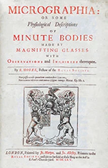
Background imageMicroscopic Collection: Title page Illustration from 'Micrographia'

Title page Illustration from 'Micrographia'
2626681 Title page Illustration from 'Micrographia'; (add.info.: Title page with Illustration from 'Micrographia' 1665; by Robert Hooke, 1635-1703)

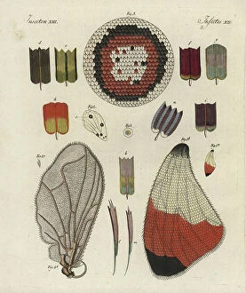
Background imageMicroscopic Collection: Butterfly wing, showing structure, skeleton, scales and microscopic detail

Butterfly wing, showing structure, skeleton, scales and microscopic detail
FLO4573972 Butterfly wing, showing structure, skeleton, scales and microscopic detail. Handcoloured copperplate engraving from Bertuch's " Bilderbuch fur Kinder"

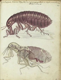
Background imageMicroscopic Collection: Human flea, (Pulex irritans), 1770-1787. Creator: Jan Brandes

Human flea, (Pulex irritans), 1770-1787. Creator: Jan Brandes
Human flea, (Pulex irritans), 1770-1787. Female flea seen through a microscope, internal organs shown. With inscription. Part of Jan Brandes sketchbook, dl. 1 (1808), p. 209

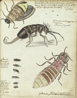
Background imageMicroscopic Collection: Javanese and Gelderland glowworms, 1770-1787. Creator: Jan Brandes

Javanese and Gelderland glowworms, 1770-1787. Creator: Jan Brandes
Javanese and Gelderland glowworms, 1770-1787. Glowworms seen under a microscope. With inscription. Part of Jan Brandes sketchbook, dl. 1 (1808), p. 213

Background imageMicroscopic Collection: Focus stacked, inverted image of Diatoms on a microscope slide

Focus stacked, inverted image of Diatoms on a microscope slide. Diatoms are single-celled algae which produce approximately 25% of the oxygen we breathe as well as being responsible for approximately

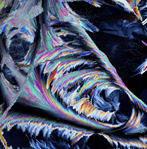
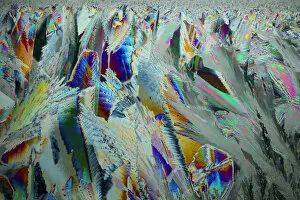
Background imageMicroscopic Collection: Sheet ice crystals viewed by polarised light

Sheet ice crystals viewed by polarised light

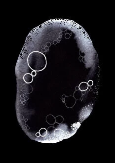
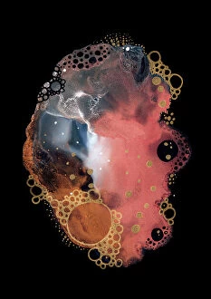
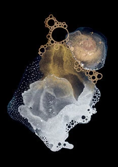
Background imageMicroscopic Collection: Specimen XII

Specimen XII
Amy Louise Baker

Background imageMicroscopic Collection: Specimen XI

Specimen XI
Amy Louise Baker

Background imageMicroscopic Collection: Specimen X

Specimen X
Amy Louise Baker

Background imageMicroscopic Collection: Specimen IX

Specimen IX
Amy Louise Baker

Background imageMicroscopic Collection: Specimen VIII

Specimen VIII
Amy Louise Baker

Background imageMicroscopic Collection: Specimen VII

Specimen VII
Amy Louise Baker

Background imageMicroscopic Collection: Specimen VI

Specimen VI
Amy Louise Baker

Background imageMicroscopic Collection: Specimen V

Specimen V
Amy Louise Baker

Background imageMicroscopic Collection: Specimen IV

Specimen IV
Amy Louise Baker

Background imageMicroscopic Collection: Specimen III

Specimen III
Amy Louise Baker

Background imageMicroscopic Collection: Specimen II

Specimen II
Amy Louise Baker

Background imageMicroscopic Collection: Specimen I

Specimen I
Amy Louise Baker

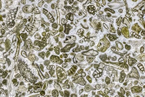
Background imageMicroscopic Collection: Limestone microfossils in a 0. 03mm-thick slice of fossil-rich limestone viewed at high

Limestone microfossils in a 0. 03mm-thick slice of fossil-rich limestone viewed at high magnification
Limestone microfossils in a 0.03mm-thick slice of fossil-rich limestone viewed at high magnification, image area is 3mm across in real life

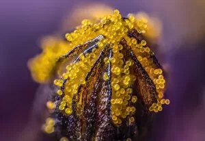
Background imageMicroscopic Collection: African daisy (Osteospermum jucundum) at approx 10x magnification

African daisy (Osteospermum jucundum) at approx 10x magnification, composite flower showing individual florets full of golden pollen grains, Monmouthshire, Wales, UK, July

Background imageMicroscopic Collection: Plankton at night, Palau, Philippine Sea

Plankton at night, Palau, Philippine Sea

Background imageMicroscopic Collection: Ice crystals viewed by polarised light

Ice crystals viewed by polarised light

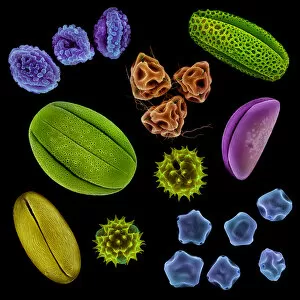
Background imageMicroscopic Collection: Various pollen grains from several species of UK plant. False coloured scanning electron micrograph

Various pollen grains from several species of UK plant. False coloured scanning electron micrograph. Digital composite

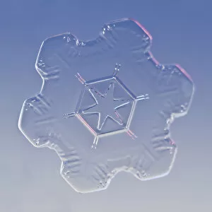
Background imageMicroscopic Collection: Snowflake magnified under microscope, Lilehammer, Norway

Snowflake magnified under microscope, Lilehammer, Norway

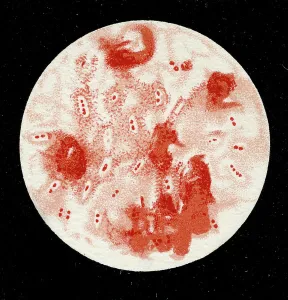
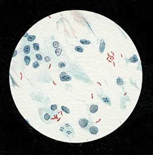
Background imageMicroscopic Collection: Colony of Haemophilus influenzae, 1906 (litho)

Colony of Haemophilus influenzae, 1906 (litho)
3501914 Colony of Haemophilus influenzae, 1906 (litho) by German School, (20th century); Private Collection; (add.info.: Bacteria constitute a large domain of prokaryotic microorganisms)

Background imageMicroscopic Collection: Colony of Mycobacterium leprae, 1906 (litho)

Colony of Mycobacterium leprae, 1906 (litho)
3501912 Colony of Mycobacterium leprae, 1906 (litho) by German School, (20th century); Private Collection; (add.info.: Bacteria constitute a large domain of prokaryotic microorganisms)

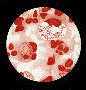
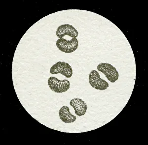
Background imageMicroscopic Collection: Colony of Micrococcus Gonorrhoea, 1906 (litho)

Colony of Micrococcus Gonorrhoea, 1906 (litho)
3501903 Colony of Micrococcus Gonorrhoea, 1906 (litho) by German School, (20th century); Private Collection; (add.info.: Bacteria constitute a large domain of prokaryotic microorganisms)

Background imageMicroscopic Collection: Colony of Streptococcus pneumoniae with and without bubble capsule, 1906 (litho)

Colony of Streptococcus pneumoniae with and without bubble capsule, 1906 (litho)
3501899 Colony of Streptococcus pneumoniae with and without bubble capsule, 1906 (litho) by German School, (20th century); Private Collection; (add.info)

Background imageMicroscopic Collection: Colony of Streptococcus Pneumoniae, 1906 (litho)

Colony of Streptococcus Pneumoniae, 1906 (litho)
3501898 Colony of Streptococcus Pneumoniae, 1906 (litho) by German School, (20th century); Private Collection; (add.info.: Bacteria constitute a large domain of prokaryotic microorganisms)

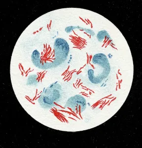
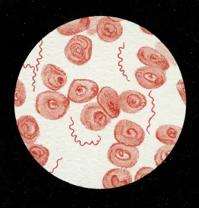
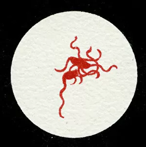
Background imageMicroscopic Collection: Spirillum of the Lyme disease causing bacteria, Spirochaetes Borrelia Recurrentis

Spirillum of the Lyme disease causing bacteria, Spirochaetes Borrelia Recurrentis, in blood sample, 1906 (litho)
3501919 Spirillum of the Lyme disease causing bacteria, Spirochaetes Borrelia Recurrentis, in blood sample, 1906 (litho) by German School

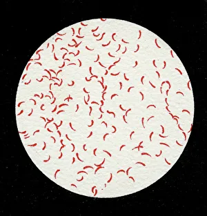
Background imageMicroscopic Collection: Colony of Vibrio cholerae, 1906 (litho)

Colony of Vibrio cholerae, 1906 (litho)
3501918 Colony of Vibrio cholerae, 1906 (litho) by German School, (20th century); Private Collection; (add.info.: Bacteria constitute a large domain of prokaryotic microorganisms)

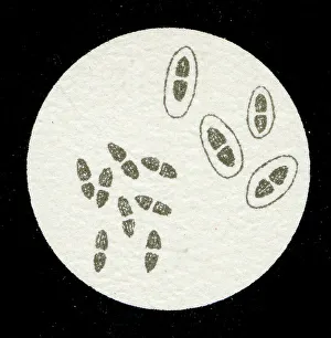
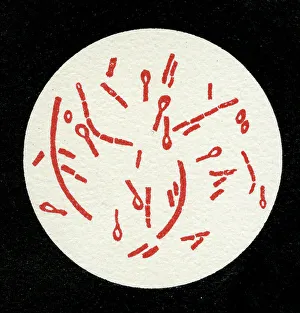
Background imageMicroscopic Collection: Colony of Clostridium tetani with spores, 1906 (litho)

Colony of Clostridium tetani with spores, 1906 (litho)
3501913 Colony of Clostridium tetani with spores, 1906 (litho) by German School, (20th century); Private Collection; (add.info.: Bacteria constitute a large domain of prokaryotic microorganisms)

Background imageMicroscopic Collection: Colony of Mycobacterium tuberculosis, 1906 (litho)

Colony of Mycobacterium tuberculosis, 1906 (litho)
3501910 Colony of Mycobacterium tuberculosis, 1906 (litho) by German School, (20th century); Private Collection; (add.info.: Bacteria constitute a large domain of prokaryotic microorganisms)

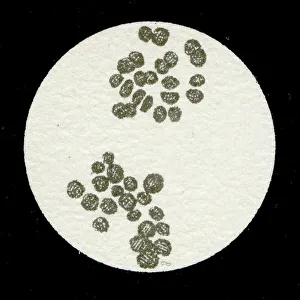
Background imageMicroscopic Collection: A colony of Staphylococcus pyogenes after cell division, 1906 (litho)

A colony of Staphylococcus pyogenes after cell division, 1906 (litho)
3501897 A colony of Staphylococcus pyogenes after cell division, 1906 (litho) by German School, (20th century); Private Collection; (add.info)

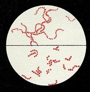
Background imageMicroscopic Collection: Colony of Streptococcus pyogenes, 1906 (litho)

Colony of Streptococcus pyogenes, 1906 (litho)
3501894 Colony of Streptococcus pyogenes, 1906 (litho) by German School, (20th century); Private Collection; (add.info.: Bacteria constitute a large domain of prokaryotic microorganisms)

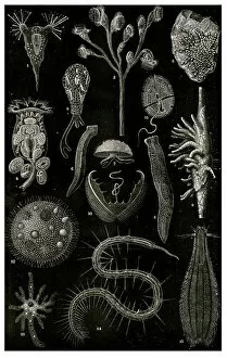
Background imageMicroscopic Collection: Varieties of microscopic Marine organisms, 1900 (litho)

Varieties of microscopic Marine organisms, 1900 (litho)
3501931 Varieties of microscopic Marine organisms, 1900 (litho) by German School, (20th century); Private Collection; Prismatic Pictures

Background imageMicroscopic Collection: Colony of Yersinia pestis, 1906 (litho)

Colony of Yersinia pestis, 1906 (litho)
3501917 Colony of Yersinia pestis, 1906 (litho) by German School, (20th century); Private Collection; (add.info.: Bacteria constitute a large domain of prokaryotic microorganisms)

Background imageMicroscopic Collection: Colony of Salmonella Typhi showing Bacilli with flagella, 1906 (litho)

Colony of Salmonella Typhi showing Bacilli with flagella, 1906 (litho)
3501916 Colony of Salmonella Typhi showing Bacilli with flagella, 1906 (litho) by German School, (20th century); Private Collection; (add.info)

Background imageMicroscopic Collection: Colony of Salmonella typhi, 1906 (litho)

Colony of Salmonella typhi, 1906 (litho)
3501915 Colony of Salmonella typhi, 1906 (litho) by German School, (20th century); Private Collection; (add.info.: Bacteria constitute a large domain of prokaryotic microorganisms)

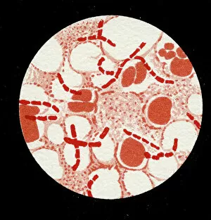
Background imageMicroscopic Collection: Colony of Bacillus Anthracis in a human spleen, 1906 (litho)

Colony of Bacillus Anthracis in a human spleen, 1906 (litho)
3501905 Colony of Bacillus Anthracis in a human spleen, 1906 (litho) by German School, (20th century); Private Collection; (add.info)


All Professionally Made to Order for Quick Shipping

"Exploring the Microscopic World: A Glimpse into Nature's Hidden Wonders" Step into a realm unseen by the naked eye, where tiny creatures and intricate structures come to life. In Picture No. 10851646, we are captivated by the Scanning Electron Micrograph (SEM) of a Praying Mantis, magnified x30. Its delicate features and mesmerizing details reveal a world within our reach. Moving on to another fascinating creature, the Tardigrade or 'Water Bear', magnified x1250 in an SEM image. This resilient micro-animal showcases its remarkable adaptability under extreme conditions. It reminds us that even in the tiniest forms of life, strength can be found. The Fruit Fly takes center stage next with an SEM image at x300 magnification. Despite its minuscule size, this common insect holds secrets waiting to be discovered under closer inspection. Its intricate body structure hints at its role in nature's grand tapestry. Diatoms from marine plankton samples grace our vision next - their elegant shapes and patterns resembling miniature works of art created by nature itself. These microscopic organisms play a vital role in Earth's ecosystems as primary producers. An illustration of a Flea C017/3435 introduces us to yet another hidden world beneath our feet - one filled with parasites and symbiotic relationships that shape entire ecosystems on both macro and micro scales. Diving deeper into marine diatoms at x25 magnification reveals their stunning beauty – intricate designs etched onto tiny shells floating through vast oceans, silently contributing to Earth's biodiversity. Anton van Leeuwenhoek's observations of Animalcules circa 1795 remind us of humanity's earliest encounters with microscopic lifeforms – pioneering discoveries that forever changed our understanding of biology and paved the way for modern scientific advancements. A Macro Photograph unveils Paenibacillus bacteria colonies thriving amidst laboratory-imposed stresses, showcasing their ability to adapt and survive in challenging environments.