Fraud Blocker Skip to main content

Microscopic Collection (page 7)

"Exploring the Microscopic World: A Glimpse into Nature's Hidden Wonders" Step into a realm unseen by the naked eye

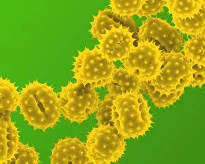
Background imageMicroscopic Collection: Picture No. 11014605

Picture No. 11014605
Grass Pollen Scanning Electron Micrograph (SEM): Magnification 2 x9410 (if print A4 size: 29.7 cm wide) - Grass pollen is wind pollinated

Background imageMicroscopic Collection: Picture No. 11014602

Picture No. 11014602
Daffodil Pollen Scanning Electron Micrograph (SEM): Magnification x16900 (if print A4 size: 29.7 cm wide) - Daffodils are popular and common springtime flowers. Insect pollinated Date:

Background imageMicroscopic Collection: Picture No. 11014603

Picture No. 11014603
Daisy Pollen Scanning Electron Micrograph (SEM): Magnification 2 x9k (if print A4 size: 29.7 cm wide) - Daisies are small insect pollinated flowers; common in lawns Date:

Background imageMicroscopic Collection: Picture No. 11014600

Picture No. 11014600
Crocus Pollen Scanning Electron Micrograph (SEM): Magnification x4000 (if print A4 size: 29.7 cm wide) - Crocus is a common spring flower, insect pollinated Date:

Background imageMicroscopic Collection: Picture No. 11014598

Picture No. 11014598
Celandine Pollen Scanning Electron Micrograph (SEM): Magnification x17, 600 (if print A4 size: 29.7 cm wide) - is a yellow springtime flower. Insect pollinated, it is toxic to humans Date:

Background imageMicroscopic Collection: Picture No. 11014597

Picture No. 11014597
Celandine Pollen Scanning Electron Micrograph (SEM): Magnification x4900 (if print A4 size: 29.7 cm wide) - is a yellow springtime flower. Insect pollinated, it is toxic to humans. Date:

Background imageMicroscopic Collection: Picture No. 11014595

Picture No. 11014595
Bumblebee Scanning Electron Micrograph (SEM): Magnification x40 (if print A4 size: 29.7 cm wide) (Bombus terrestris) Date:

Background imageMicroscopic Collection: Picture No. 10873599

Picture No. 10873599
Human Crab Louse (Phthirus pubis) Date:

Background imageMicroscopic Collection: Picture No. 10877002

Picture No. 10877002
Scanning Electron Micrograph (SEM): Cat hairs Date:

Background imageMicroscopic Collection: Picture No. 10876987

Picture No. 10876987
Scanning Electron Micrograph (SEM): Human skin section across vein showing red blood cells Date:

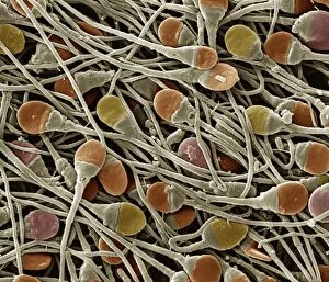
Background imageMicroscopic Collection: Picture No. 10876990

Picture No. 10876990
Scanning Electron Micrograph (SEM): Human sperm Date:

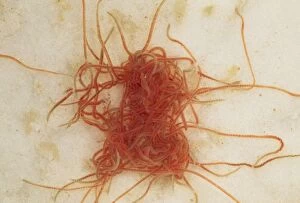
Background imageMicroscopic Collection: Picture No. 10855792

Picture No. 10855792
Tubifex Worms - freshwater stream (Lumbriculus variegatus ) Date:

Background imageMicroscopic Collection: Radiolaria models

Radiolaria models
Models of two radiolaria made in papier mache by Vaclav Fric

Background imageMicroscopic Collection: Marine Diatoms

Marine Diatoms
LRMC-3 Scanning Electron Micrograph (SEM): Marine Diatoms Magnification x2400 (when printed A4, 29.7 cm cm wide) David McCarthy and Dae Sasitorn / ardea.com Last Refuge contact details

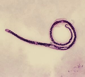
Background imageMicroscopic Collection: Light Micrograph (SEM): Micro-filarial worm - Magnification x 3000 (if print A4 size: 29. 7 cm wide)

Light Micrograph (SEM): Micro-filarial worm - Magnification x 3000 (if print A4 size: 29. 7 cm wide)
LRDS-314 Light Micrograph (SEM): Micro-filarial worm Magnification x 3000 (if print A4 size: 29.7 cm wide) Wuchereria bancrofti Elephantiasis David Spears (Last Refuge)

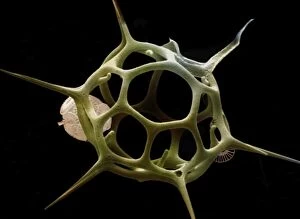
Background imageMicroscopic Collection: Silicoflagellate

Silicoflagellate
LRDS-297 Scanning Electron Micrograph (SEM): Silicoflagellate, Skeleton bearing stage of Dictyocha speculum Magnification x3

Background imageMicroscopic Collection: Euglena gracilis

Euglena gracilis protists. Light micrograph of a group of Euglena gracilis protists. These organisms possess a unique combination of plant and animal characters. Like the algae, E

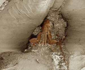
Background imageMicroscopic Collection: Smallest book and millipede, SEM

Smallest book and millipede, SEM
Smallest book in the world with a millipede. Coloured scanning electron micrograph (SEM) of a millipede (class Diplopoda) and the smallest published book in the world

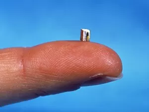
Background imageMicroscopic Collection: Smallest book

Smallest book in the world held on a human finger. This is the smallest published book in the world, a tiny leather-bound volume that measures just 2.4 by 2.9 millimetres in page area

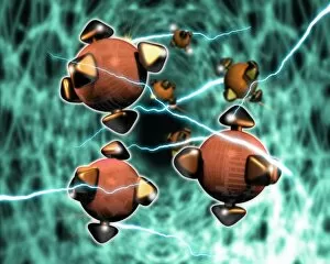
Background imageMicroscopic Collection: Nanorobots

Nanorobots. Computer artwork of nanorobots in the brain, surrounded by nerve impulses. Microscopic robot technology could be developed in the future to treat diseases in new ways

Background imageMicroscopic Collection: ROBERT HOOKE 1635 - 1703

ROBERT HOOKE 1635 - 1703
ROBERT HOOKE English scientist. Author of Micrographia (1665), in which he published results of his microscopic investigations

Background imageMicroscopic Collection: Microscopic Fish Gill

Microscopic Fish Gill
PM-1947 Fish Anatomy - Fish gill under microscope. Pat Morris Please note that prints are for personal display purposes only and may not be reproduced in anyway

Background imageMicroscopic Collection: Scanning Electron Micrograph (SEM): Mosquito, Magnification x 35 (A4 size: 29. 7 cm width)

Scanning Electron Micrograph (SEM): Mosquito, Magnification x 35 (A4 size: 29. 7 cm width)
LRDS-84 Mosquito Scanning Electron Micrograph (SEM) Culex pipiens Magnification x 35 (A4 size: 29.7 cm width) Coloured by hand to enhance natural features

Background imageMicroscopic Collection: Light Micrograph : Flattened Mayfly Nymph, Magnification x 10 (A4 size: 29. 7 cm width)

Light Micrograph : Flattened Mayfly Nymph, Magnification x 10 (A4 size: 29. 7 cm width)
LRDS-80 Flattened Mayfly Nymph Light Micrograph Ecdyonurus venosus Magnification x 10 (A4 size: 29.7 cm width) Credit: David Spears (last refuge)

Background imageMicroscopic Collection: Scanning Electron Micrograph (SEM): Flour Mite, Magnification x 500 (A4 size: 29. 7 cm width)

Scanning Electron Micrograph (SEM): Flour Mite, Magnification x 500 (A4 size: 29. 7 cm width)
LRDS-58 Flour Mite Scanning Electron Micrograph (SEM) Acarus siro Magnification x 500 (A4 size: 29.7 cm width) Coloured by hand to enhance natural features

Background imageMicroscopic Collection: Scanning Electron Micrograph (SEM): Cat Flea, Magnification x250 (A4 size: 29. 7 cm width)

Scanning Electron Micrograph (SEM): Cat Flea, Magnification x250 (A4 size: 29. 7 cm width)
LRDS-54 Cat Flea Scanning Electron Micrograph (SEM) Ctenocephalides felis Magnification x250 (A4 size: 29.7 cm width) Coloured by hand to enhance natural features

Background imageMicroscopic Collection: Light Micrograph (LM): Transverse section of Dehiscence Lilium Anthers with Pollen; Magnification

Light Micrograph (LM): Transverse section of Dehiscence Lilium Anthers with Pollen; Magnification x300 (on 10)
LRDS-200 Light Micrograph (LM): Transverse section of Dehiscence Lilium Anthers with Pollen Magnification x300 (on 10.5 cm width print) David Spears (Last Refuge)

Background imageMicroscopic Collection: Scanning Electron Micrograph (SEM): Chigoe Flea, Magnification x 200 (A4 size: 29. 7 cm width)

Scanning Electron Micrograph (SEM): Chigoe Flea, Magnification x 200 (A4 size: 29. 7 cm width)
LRDS-137 Chigoe Flea Scanning Electron Micrograph (SEM) Tunga penetrans Magnification x 200 (A4 size: 29.7 cm width) Coloured by hand to enhance natural features

Background imageMicroscopic Collection: Scanning Electron Micrograph (SEM): Springtail, Order Collembola; Magnification x 150 (A4 size)

Scanning Electron Micrograph (SEM): Springtail, Order Collembola; Magnification x 150 (A4 size: 29. 7 cm width)
LRDS-122 Springtail, Order Collembola Scanning Electron Micrograph (SEM) Magnification x 150 (A4 size: 29.7 cm width) Coloured by hand to enhance natural features

Background imageMicroscopic Collection: nanotechnology

nanotechnology
biotechnology fighting disease

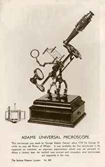
Background imageMicroscopic Collection: Adams Universal Microscope

Adams Universal Microscope - made by George Adams Snr. about 1755 for King George III, while he was still Prince of Wales

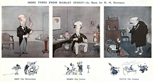
Background imageMicroscopic Collection: More Types From Harley Street by H M Bateman

More Types From Harley Street by H M Bateman. Skin - The Microscopic. The one with the magnifying glass and the test tubes. Brain - The Genius. He knows all about the mind. Nerve - The Intense

Background imageMicroscopic Collection: X-Ray - Microscopic x-ray view of a small head louse

X-Ray - Microscopic x-ray view of a small head louse. Part of Box 165 Boswell Collection - X-Rays. Date: circa 1900

Background imageMicroscopic Collection: Binocular microscope large Best. London, around 1890

Binocular microscope large Best. London, around 1890
Binocular microscope large Best. Signed: R. & J. Beck Ltd London 19901. London, around 1890. The Large Best microscope was the top product of R. and J

Background imageMicroscopic Collection: Changeable cercaria, Cercaria mutabilis

Changeable cercaria, Cercaria mutabilis.. Handcolored copperplate zoological engraving from George Shaw and Frederick Nodders The Naturalists Miscellany, 1792

Background imageMicroscopic Collection: Common moss, Phascum cuspidatum, and pond algae

Common moss, Phascum cuspidatum, and pond algae.. Magnified image of common moss, Phascum cuspidatum 1, and microscopic enlargment of pond algae, Hydrodictyon utriculatum 2

Background imageMicroscopic Collection: POLIOVIRUS, 1953. An early image of the poliovirus taken by an electron microscope

POLIOVIRUS, 1953. An early image of the poliovirus taken by an electron microscope. Photograph, 1953


All Professionally Made to Order for Quick Shipping

"Exploring the Microscopic World: A Glimpse into Nature's Hidden Wonders" Step into a realm unseen by the naked eye, where tiny creatures and intricate structures come to life. In Picture No. 10851646, we are captivated by the Scanning Electron Micrograph (SEM) of a Praying Mantis, magnified x30. Its delicate features and mesmerizing details reveal a world within our reach. Moving on to another fascinating creature, the Tardigrade or 'Water Bear', magnified x1250 in an SEM image. This resilient micro-animal showcases its remarkable adaptability under extreme conditions. It reminds us that even in the tiniest forms of life, strength can be found. The Fruit Fly takes center stage next with an SEM image at x300 magnification. Despite its minuscule size, this common insect holds secrets waiting to be discovered under closer inspection. Its intricate body structure hints at its role in nature's grand tapestry. Diatoms from marine plankton samples grace our vision next - their elegant shapes and patterns resembling miniature works of art created by nature itself. These microscopic organisms play a vital role in Earth's ecosystems as primary producers. An illustration of a Flea C017/3435 introduces us to yet another hidden world beneath our feet - one filled with parasites and symbiotic relationships that shape entire ecosystems on both macro and micro scales. Diving deeper into marine diatoms at x25 magnification reveals their stunning beauty – intricate designs etched onto tiny shells floating through vast oceans, silently contributing to Earth's biodiversity. Anton van Leeuwenhoek's observations of Animalcules circa 1795 remind us of humanity's earliest encounters with microscopic lifeforms – pioneering discoveries that forever changed our understanding of biology and paved the way for modern scientific advancements. A Macro Photograph unveils Paenibacillus bacteria colonies thriving amidst laboratory-imposed stresses, showcasing their ability to adapt and survive in challenging environments.