Fraud Blocker Skip to main content

Microscopic Collection (page 5)

"Exploring the Microscopic World: A Glimpse into Nature's Hidden Wonders" Step into a realm unseen by the naked eye

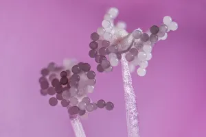
Background imageMicroscopic Collection: Hollyhock (Alcea rosea), pollen grains on anthers, aprox 12x magnification, Monmouthshire

Hollyhock (Alcea rosea), pollen grains on anthers, aprox 12x magnification, Monmouthshire, Wales, UK

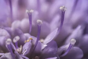
Background imageMicroscopic Collection: Small scabious (Scabiosa columbaria) with three tiny (1mm) Thrips (Order Thysanoptera

Small scabious (Scabiosa columbaria) with three tiny (1mm) Thrips (Order Thysanoptera) on a petal, Monmouthshire, Wales, UK

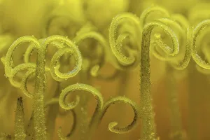
Background imageMicroscopic Collection: Dandelion (Taraxacum officinale) stigma open to receive pollen for fertilization

Dandelion (Taraxacum officinale) stigma open to receive pollen for fertilization, Monmouthshire, Wales, UK, July

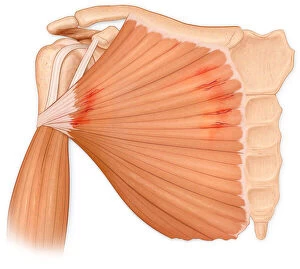
Background imageMicroscopic Collection: Microscopic muscle fiber tears of the pectoralis major muscle

Microscopic muscle fiber tears of the pectoralis major muscle
Artwork, Bone, Clavicle, Human Anatomy, humerus, Illustration, inflammation, Injury, Medical, Microscopic, Muscle, muscle fiber tear, Pectoralis major muscle, Pectoralis Major Tear Syndrome, scapula

Background imageMicroscopic Collection: PT_0241

PT_0241
Cotyledon orbiculata Plant Foliage Growing Close-up View - Front view View - Side view Orientation - Vertical orientation One Creative Abstract Microscopic Contrasting - Colour Textured Unusual plant

Background imageMicroscopic Collection: Dahlia, abstract deep red pattern

Dahlia, abstract deep red pattern against black

Background imageMicroscopic Collection: Picture No. 12479490

Picture No. 12479490
Marine fish larvae eat microplastics. Small pieces Date:

Background imageMicroscopic Collection: Picture No. 12479491

Picture No. 12479491
Marine fish larvae eat microplastics. Small pieces Date:

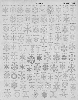
Background imageMicroscopic Collection: Snow, early 19th century. Creator: J Moffat

Snow, early 19th century. Creator: J Moffat
Snow, early 19th century. Illustrations showing the structure of ice crystals which form snowflakes, as seen under the microscope

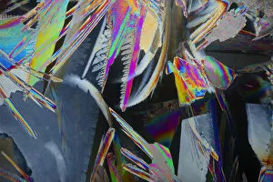
Background imageMicroscopic Collection: Ice crystals viewed by polarised light

Ice crystals viewed by polarised light

Background imageMicroscopic Collection: Fossil Wood in Sandstone, from a Moraine in Latitude 85°South, c1908, (1909)

Fossil Wood in Sandstone, from a Moraine in Latitude 85°South, c1908, (1909)
Fossil Wood in Sandstone, from a Moraine in Latitude 85° South, c1908, (1909). Microscope sample from glacial debris in Antarctica

Background imageMicroscopic Collection: Ice Crystals in Crevasse, June-July 1911, (1913). Artist: Charless Wright

Ice Crystals in Crevasse, June-July 1911, (1913). Artist: Charless Wright
Ice Crystals in Crevasse, June-July 1911, (1913). Microscope views showing ther formation of ice. Expedition physicist Charles Wright carried out experiments

Background imageMicroscopic Collection: Microscopes and microscopical objects, 1750

Microscopes and microscopical objects, 1750. I: Wilsons pocket microscope. II: Scroll microscope. III: Tripod microscope - improved form of Marshalls double microscope

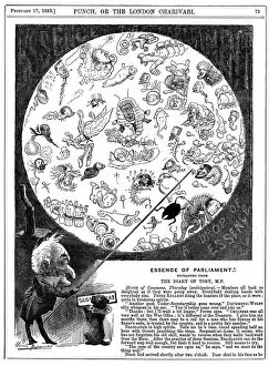
Background imageMicroscopic Collection: Essence of Parliament, 1883. Artist: Harry Furniss

Essence of Parliament, 1883. Artist: Harry Furniss
Essence of Parliament, 1883. Cartoon marking the opening of the first session of Parliament for 1883, showing the politicians as microbes in a drop of water

Background imageMicroscopic Collection: Microscopic view of animal cell

Microscopic view of animal cell

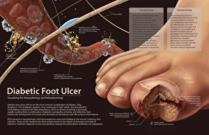
Background imageMicroscopic Collection: Histopathology and pathophysiology of diabetic food ulcers

Histopathology and pathophysiology of diabetic food ulcers

Background imageMicroscopic Collection: Picture No. 11014634

Picture No. 11014634
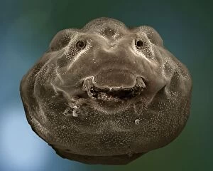
Common Frog - mid stage of tadpole development Scanning Electron Micrograph (SEM): Magnification x 85 (if print A4 size: 29.7 cm wide) - Gills are resorbed and jaws better developed

Background imageMicroscopic Collection: Picture No. 11014629

Picture No. 11014629
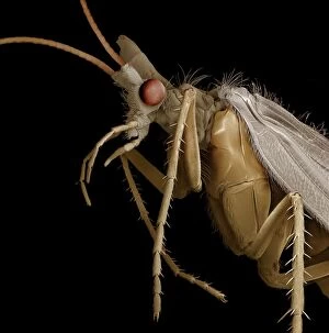
Caddis Fly Scanning Electron Micrograph (SEM): Magnification x 20 (if print A4 size: 29.7 cm wide) - Caddis flies spend most of their lives as aquatic larvae in ponds and streams

Background imageMicroscopic Collection: Picture No. 11014630

Picture No. 11014630
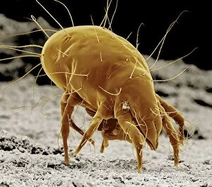
Cheese Mite Scanning Electron Micrograph (SEM): Magnification x 350 (if print A4 size: 29.7 cm wide) (Tyrolichus casei) Date:

Background imageMicroscopic Collection: Picture No. 11014628

Picture No. 11014628
Caenorhabditis elegans Scanning Electron Micrograph (SEM): Magnification x 1750 (if print A4 size: 29.7 cm wide) - This tiny free living nematode worm is extensively used in medical

Background imageMicroscopic Collection: Picture No. 11014624

Picture No. 11014624
Escherishia Coli Bacteria / E. Coli - affected by antibiotic Scanning Electron Micrograph (SEM): Magnification x25, 000 (if print A4 size)

Background imageMicroscopic Collection: Picture No. 11014620

Picture No. 11014620
Mosquito Larvae Scanning Electron Micrograph (SEM): Magnification x40 (if print A4 size: 29.7 cm wide) - The malarial mosquito larvae live in pools and puddles; almost any standing water will do

Background imageMicroscopic Collection: Picture No. 11014623

Picture No. 11014623
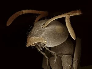
Black Garden Ant Scanning Electron Micrograph (SEM): Magnification x120 (if print A4 size: 29.7 cm wide) (Lasius niger) Date:

Background imageMicroscopic Collection: Picture No. 11014622

Picture No. 11014622
Mosquito Scanning Electron Micrograph (SEM): Magnification x90 (if print A4 size: 29.7 cm wide) - Portrait of the malarial mosquito showing the antenae and palps

Background imageMicroscopic Collection: Picture No. 11014616

Picture No. 11014616
Sunflower Pollen Scanning Electron Micrograph (SEM): Magnification x15, 000 (if print A4 size: 29.7 cm wide) - Sunflowers have huge inflorescences that shed large amounts of pollen that is carried

Background imageMicroscopic Collection: Picture No. 11014592

Picture No. 11014592
Aconite Anther - with pollen Scanning Electron Micrograph (SEM): Magnification x 11150 (if print A4 size: 29.7 cm wide) - Aconites are attractive springtime garden flowers - They are insect

Background imageMicroscopic Collection: Picture No. 11014591

Picture No. 11014591
Aconite Anther - with pollen Scanning Electron Micrograph (SEM): Magnification x 450 (if print A4 size: 29.7 cm wide) - Aconites are attractive springtime garden flowers - They are insect pollinated

Background imageMicroscopic Collection: Picture No. 10873637

Picture No. 10873637
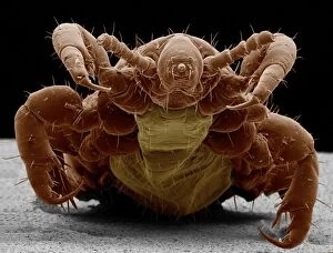
Human Head Louse (Pediculus humanus capitis) Date:

Background imageMicroscopic Collection: Colonial Green Algae SPH 2999 Volvox sp. © Steve Hopkin / ardea. com

Colonial Green Algae SPH 2999 Volvox sp. © Steve Hopkin / ardea. com
SPH-2999 Colonial Green ALGAE Volvox sp. Steve Hopkin Microscopic contact details: prints@ardea.com tel: +44 (0) 20 8318 1401

Background imageMicroscopic Collection: This is a microscopic look at the detail on an Indian feather duster worm (Sabellastarte indica)

This is a microscopic look at the detail on an Indian feather duster worm (Sabellastarte indica) one of the larger of this family reaching 4 inches across, Yap, Micronesia

Background imageMicroscopic Collection: An intense and continuing interest in all aspects of the plot to assassinate Abraham Lincoln

An intense and continuing interest in all aspects of the plot to assassinate Abraham Lincoln resulted in curiosities like this, even though it was not published until 1867, two years after the event

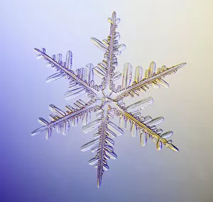
Background imageMicroscopic Collection: Photo-microscope view of a real snowflake showing the classic 6-sided star shape

Photo-microscope view of a real snowflake showing the classic 6-sided star shape

Background imageMicroscopic Collection: Photomicroscopic Close Up Of Two Snowflake Crystals, Alaska

Photomicroscopic Close Up Of Two Snowflake Crystals, Alaska

Background imageMicroscopic Collection: Photomicroscopic Close Up Of A Snowflake Crystal, Alaska

Photomicroscopic Close Up Of A Snowflake Crystal, Alaska

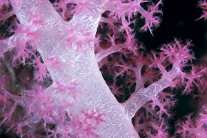
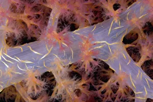
Background imageMicroscopic Collection: Palau, Alcyonarian Coral Pink Detail, Black Background

Palau, Alcyonarian Coral Pink Detail, Black Background

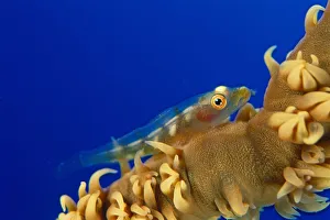
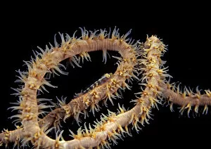
Background imageMicroscopic Collection: Hawaii, Goby (Bryaninops Yongei) On Wire Coral (Cirrhipathes Anguina) Blue Background

Hawaii, Goby (Bryaninops Yongei) On Wire Coral (Cirrhipathes Anguina) Blue Background

Background imageMicroscopic Collection: Solomon Islands, Bright Pink Soft Coral With Goby Atop, Close-Up Detail

Solomon Islands, Bright Pink Soft Coral With Goby Atop, Close-Up Detail

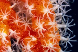
Background imageMicroscopic Collection: Micronesia, Sponge Covered Gorgonian Coral Showing Polyp Detail, Orange And White

Micronesia, Sponge Covered Gorgonian Coral Showing Polyp Detail, Orange And White

Background imageMicroscopic Collection: Fiji, Close-Up Of Soft Coral Showing Polyps Feeding (Siphonogorgia Sp?)

Fiji, Close-Up Of Soft Coral Showing Polyps Feeding (Siphonogorgia Sp?)

Background imageMicroscopic Collection: Solomon Islands, Goby On Sea Whip, Close-Up Detail A90A

Solomon Islands, Goby On Sea Whip, Close-Up Detail A90A

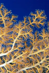
Background imageMicroscopic Collection: Indonesia, Eggs Can Be Seen Through The Translucent Flesh Of This Alcyonarian Coral (Dendronephthya)

Indonesia, Eggs Can Be Seen Through The Translucent Flesh Of This Alcyonarian Coral (Dendronephthya)

Background imageMicroscopic Collection: USA, Goby (Bryaninops yongei) at night on feeding wire coral (Cirrhipathes anguina)

USA, Goby (Bryaninops yongei) at night on feeding wire coral (Cirrhipathes anguina) in Pacific ocean; Hawaii

Background imageMicroscopic Collection: Photo-microscope view of a real snowflake showing the classic 6-sided star shape

Photo-microscope view of a real snowflake showing the classic 6-sided star shape


All Professionally Made to Order for Quick Shipping

"Exploring the Microscopic World: A Glimpse into Nature's Hidden Wonders" Step into a realm unseen by the naked eye, where tiny creatures and intricate structures come to life. In Picture No. 10851646, we are captivated by the Scanning Electron Micrograph (SEM) of a Praying Mantis, magnified x30. Its delicate features and mesmerizing details reveal a world within our reach. Moving on to another fascinating creature, the Tardigrade or 'Water Bear', magnified x1250 in an SEM image. This resilient micro-animal showcases its remarkable adaptability under extreme conditions. It reminds us that even in the tiniest forms of life, strength can be found. The Fruit Fly takes center stage next with an SEM image at x300 magnification. Despite its minuscule size, this common insect holds secrets waiting to be discovered under closer inspection. Its intricate body structure hints at its role in nature's grand tapestry. Diatoms from marine plankton samples grace our vision next - their elegant shapes and patterns resembling miniature works of art created by nature itself. These microscopic organisms play a vital role in Earth's ecosystems as primary producers. An illustration of a Flea C017/3435 introduces us to yet another hidden world beneath our feet - one filled with parasites and symbiotic relationships that shape entire ecosystems on both macro and micro scales. Diving deeper into marine diatoms at x25 magnification reveals their stunning beauty – intricate designs etched onto tiny shells floating through vast oceans, silently contributing to Earth's biodiversity. Anton van Leeuwenhoek's observations of Animalcules circa 1795 remind us of humanity's earliest encounters with microscopic lifeforms – pioneering discoveries that forever changed our understanding of biology and paved the way for modern scientific advancements. A Macro Photograph unveils Paenibacillus bacteria colonies thriving amidst laboratory-imposed stresses, showcasing their ability to adapt and survive in challenging environments.