Fraud Blocker Skip to main content

Microscopic Collection (page 6)

"Exploring the Microscopic World: A Glimpse into Nature's Hidden Wonders" Step into a realm unseen by the naked eye

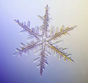
Background imageMicroscopic Collection: Photo-microscope view of a real snowflake showing the classic 6-sided star shape

Photo-microscope view of a real snowflake showing the classic 6-sided star shape

Background imageMicroscopic Collection: Photomicroscopic Close Up Of Two Snowflake Crystals, Alaska

Photomicroscopic Close Up Of Two Snowflake Crystals, Alaska

Background imageMicroscopic Collection: Photomicroscopic Close Up Of A Snowflake Crystal, Alaska

Photomicroscopic Close Up Of A Snowflake Crystal, Alaska

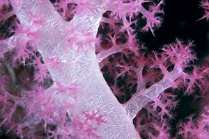
Background imageMicroscopic Collection: Palau, Alcyonarian Coral Pink Detail, Black Background

Palau, Alcyonarian Coral Pink Detail, Black Background

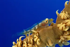
Background imageMicroscopic Collection: Hawaii, Goby (Bryaninops Yongei) On Wire Coral (Cirrhipathes Anguina) Blue Background

Hawaii, Goby (Bryaninops Yongei) On Wire Coral (Cirrhipathes Anguina) Blue Background

Background imageMicroscopic Collection: Solomon Islands, Bright Pink Soft Coral With Goby Atop, Close-Up Detail

Solomon Islands, Bright Pink Soft Coral With Goby Atop, Close-Up Detail

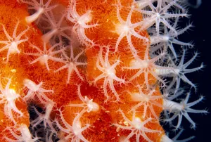
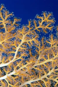
Background imageMicroscopic Collection: Micronesia, Sponge Covered Gorgonian Coral Showing Polyp Detail, Orange And White

Micronesia, Sponge Covered Gorgonian Coral Showing Polyp Detail, Orange And White

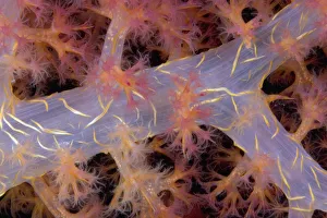
Background imageMicroscopic Collection: Fiji, Close-Up Of Soft Coral Showing Polyps Feeding (Siphonogorgia Sp?)

Fiji, Close-Up Of Soft Coral Showing Polyps Feeding (Siphonogorgia Sp?)

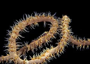
Background imageMicroscopic Collection: Solomon Islands, Goby On Sea Whip, Close-Up Detail A90A

Solomon Islands, Goby On Sea Whip, Close-Up Detail A90A

Background imageMicroscopic Collection: Indonesia, Eggs Can Be Seen Through The Translucent Flesh Of This Alcyonarian Coral (Dendronephthya)

Indonesia, Eggs Can Be Seen Through The Translucent Flesh Of This Alcyonarian Coral (Dendronephthya)

Background imageMicroscopic Collection: USA, Goby (Bryaninops yongei) at night on feeding wire coral (Cirrhipathes anguina)

USA, Goby (Bryaninops yongei) at night on feeding wire coral (Cirrhipathes anguina) in Pacific ocean; Hawaii

Background imageMicroscopic Collection: Photo-microscope view of a real snowflake showing the classic 6-sided star shape

Photo-microscope view of a real snowflake showing the classic 6-sided star shape

Background imageMicroscopic Collection: Picture No. 11675563

Picture No. 11675563
Light Micrograph (LM): Rhizopus sporangia. Date:

Background imageMicroscopic Collection: Conceptual image of platelet, red blood cell and white blood cell

Conceptual image of platelet, red blood cell and white blood cell

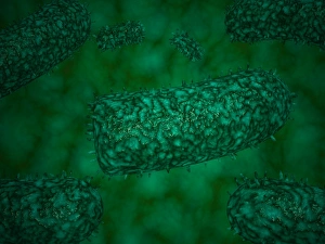
Background imageMicroscopic Collection: Stylized rabies virus particles

Stylized rabies virus particles, the cause of the viral neuroinvasive disease acute encephalitis

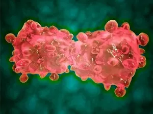
Background imageMicroscopic Collection: Microscopic view of a leukemia cell

Microscopic view of a leukemia cell

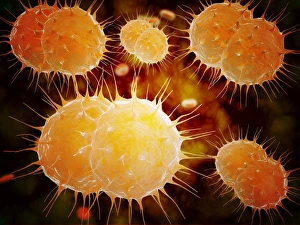
Background imageMicroscopic Collection: Microscopic view of Neisseria gonorrhoeae

Microscopic view of Neisseria gonorrhoeae. Neisseria gonorrhoeae typically infects the mucous membranes causing infections such as urethritis, cervicitis, salpingitis, pelvic inflammatory disease

Background imageMicroscopic Collection: Microscopic view of yellow fever virus

Microscopic view of yellow fever virus. Yellow fever is an acute viral disease

Background imageMicroscopic Collection: Microscopic view of Giardiasis

Microscopic view of Giardiasis, an infectious disease caused by a unicellular parasite known as Giardia lamblia

Background imageMicroscopic Collection: Microscopic view of bacterial pneumonia

Microscopic view of bacterial pneumonia. Bacterial pneumonia is a type of pneumonia caused by bacterial infection. Pneumonia can be generally defined as inflammation of the lung parenchyma

Background imageMicroscopic Collection: Microscopic view of Streptococcus pneumoniae

Microscopic view of Streptococcus pneumoniae, also known as pneumococcus. Pneumococcus is a Gram-positive coccus shaped pathogenic bacteria which causes many types of pneumococcal infections in

Background imageMicroscopic Collection: Different types of acne, non-inflammatory and inflammatory

Different types of acne, non-inflammatory and inflammatory

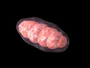
Background imageMicroscopic Collection: Conceptual image of mitochondria

Conceptual image of mitochondria

Background imageMicroscopic Collection: Microscopic view of Stachybotrys chartarum

Microscopic view of Stachybotrys chartarum, a black mold

Background imageMicroscopic Collection: Anatomy of foot fungus with microscopic close-up

Anatomy of foot fungus with microscopic close-up

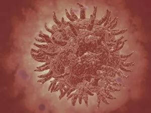
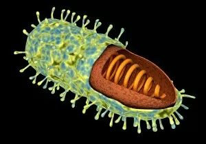
Background imageMicroscopic Collection: Conceptual image of rabies virus

Conceptual image of rabies virus

Background imageMicroscopic Collection: Picture No. 11014633

Picture No. 11014633
Penicillin Colony This is a culture of the original strain of bacteria used by Flemming in the early research into the first antibiotics. Obliquely lit macro image from a time-lapse sequence. Date:

Background imageMicroscopic Collection: Picture No. 11014631

Picture No. 11014631
Diatom Scanning Electron Micrograph (SEM): Magnification x5275 (if print A4 size: 29.7 cm wide) - The diatoms are single celled algae

Background imageMicroscopic Collection: Picture No. 11014632

Picture No. 11014632
Diatom Scanning Electron Micrograph (SEM): Magnification x 4370 (if print A4 size: 29.7 cm wide) - The diatoms are single celled algae

Background imageMicroscopic Collection: Picture No. 11014627

Picture No. 11014627
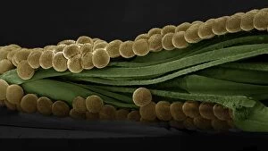
Spectacle Moth - eggs Scanning Electron Micrograph (SEM): Magnification x 35 (if print A4 size: 29.7 cm wide) (Abrostola tripartita) Date:

Background imageMicroscopic Collection: Picture No. 11014626

Picture No. 11014626
Bumblebee Scanning Electron Micrograph (SEM): Magnification x30 (if print A4 size: 29.7 cm wide) (Bombus terrestris) Date:

Background imageMicroscopic Collection: Picture No. 11014625

Picture No. 11014625
Bedbug Scanning Electron Micrograph (SEM): Magnification x120 (if print A4 size: 29.7 cm wide) (Cimex lectularius) Date:

Background imageMicroscopic Collection: Picture No. 11014621

Picture No. 11014621
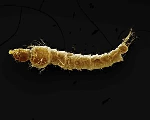
Mosquito Larvae Scanning Electron Micrograph (SEM): Magnification x40 (if print A4 size: 29.7 cm wide) - The malarial mosquito larvae live in pools and puddles; almost any standing water will do

Background imageMicroscopic Collection: Picture No. 11014619

Picture No. 11014619
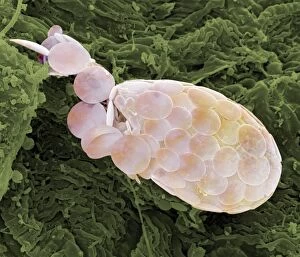
Shelled Amoeba Scanning Electron Micrograph (SEM)): Magnification x5130 (if print A4 size: 29.7 cm wide) - Testate amoebae are found in damp soil, leaf litter and pools

Background imageMicroscopic Collection: Picture No. 11014611

Picture No. 11014611
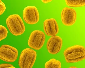
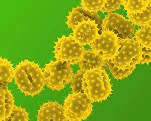
Primrose Pollen Scanning Electron Micrograph (SEM): Magnification 2 x28, 000 (if print A4 size: 29.7 cm wide) - Primroses appear in early spring and are usually yellow

Background imageMicroscopic Collection: Picture No. 11014610

Picture No. 11014610
Primrose Pollen Scanning Electron Micrograph (SEM): Magnification x12, 000 (if print A4 size: 29.7 cm wide) - Primroses appear in early spring and are usually yellow. They are insect pollinated Date:

Background imageMicroscopic Collection: Picture No. 11014609

Picture No. 11014609
Hazel Pollen Scanning Electron Micrograph (SEM): Magnification x12, 000 b1 (if print A4 size: 29.7 cm wide) - Hazel pollen is very small and is carried by the wind to stigma of other trees

Background imageMicroscopic Collection: Picture No. 11014608

Picture No. 11014608
Grass Pollen Scanning Electron Micrograph (SEM): Magnification 2 x4560 (if print A4 size: 29.7 cm wide) - Gypsophyla is a small pink or white flower; insect pollinated Date:

Background imageMicroscopic Collection: Picture No. 11014605

Picture No. 11014605
Grass Pollen Scanning Electron Micrograph (SEM): Magnification 2 x9410 (if print A4 size: 29.7 cm wide) - Grass pollen is wind pollinated

Background imageMicroscopic Collection: Picture No. 11014602

Picture No. 11014602
Daffodil Pollen Scanning Electron Micrograph (SEM): Magnification x16900 (if print A4 size: 29.7 cm wide) - Daffodils are popular and common springtime flowers. Insect pollinated Date:

Background imageMicroscopic Collection: Picture No. 11014603

Picture No. 11014603
Daisy Pollen Scanning Electron Micrograph (SEM): Magnification 2 x9k (if print A4 size: 29.7 cm wide) - Daisies are small insect pollinated flowers; common in lawns Date:

Background imageMicroscopic Collection: Picture No. 11014600

Picture No. 11014600
Crocus Pollen Scanning Electron Micrograph (SEM): Magnification x4000 (if print A4 size: 29.7 cm wide) - Crocus is a common spring flower, insect pollinated Date:

Background imageMicroscopic Collection: Picture No. 11014598

Picture No. 11014598
Celandine Pollen Scanning Electron Micrograph (SEM): Magnification x17, 600 (if print A4 size: 29.7 cm wide) - is a yellow springtime flower. Insect pollinated, it is toxic to humans Date:

Background imageMicroscopic Collection: Picture No. 11014597

Picture No. 11014597
Celandine Pollen Scanning Electron Micrograph (SEM): Magnification x4900 (if print A4 size: 29.7 cm wide) - is a yellow springtime flower. Insect pollinated, it is toxic to humans. Date:

Background imageMicroscopic Collection: Picture No. 11014595

Picture No. 11014595
Bumblebee Scanning Electron Micrograph (SEM): Magnification x40 (if print A4 size: 29.7 cm wide) (Bombus terrestris) Date:

Background imageMicroscopic Collection: Picture No. 10873599

Picture No. 10873599
Human Crab Louse (Phthirus pubis) Date:

Background imageMicroscopic Collection: Picture No. 10877002

Picture No. 10877002
Scanning Electron Micrograph (SEM): Cat hairs Date:

Background imageMicroscopic Collection: Picture No. 10876987

Picture No. 10876987
Scanning Electron Micrograph (SEM): Human skin section across vein showing red blood cells Date:


All Professionally Made to Order for Quick Shipping

"Exploring the Microscopic World: A Glimpse into Nature's Hidden Wonders" Step into a realm unseen by the naked eye, where tiny creatures and intricate structures come to life. In Picture No. 10851646, we are captivated by the Scanning Electron Micrograph (SEM) of a Praying Mantis, magnified x30. Its delicate features and mesmerizing details reveal a world within our reach. Moving on to another fascinating creature, the Tardigrade or 'Water Bear', magnified x1250 in an SEM image. This resilient micro-animal showcases its remarkable adaptability under extreme conditions. It reminds us that even in the tiniest forms of life, strength can be found. The Fruit Fly takes center stage next with an SEM image at x300 magnification. Despite its minuscule size, this common insect holds secrets waiting to be discovered under closer inspection. Its intricate body structure hints at its role in nature's grand tapestry. Diatoms from marine plankton samples grace our vision next - their elegant shapes and patterns resembling miniature works of art created by nature itself. These microscopic organisms play a vital role in Earth's ecosystems as primary producers. An illustration of a Flea C017/3435 introduces us to yet another hidden world beneath our feet - one filled with parasites and symbiotic relationships that shape entire ecosystems on both macro and micro scales. Diving deeper into marine diatoms at x25 magnification reveals their stunning beauty – intricate designs etched onto tiny shells floating through vast oceans, silently contributing to Earth's biodiversity. Anton van Leeuwenhoek's observations of Animalcules circa 1795 remind us of humanity's earliest encounters with microscopic lifeforms – pioneering discoveries that forever changed our understanding of biology and paved the way for modern scientific advancements. A Macro Photograph unveils Paenibacillus bacteria colonies thriving amidst laboratory-imposed stresses, showcasing their ability to adapt and survive in challenging environments.