
Peptide Collection
Peptides: Unveiling the Intricate World of Molecular Marvels DNA transcription and molecular model: Unlocking the secrets of life


All Professionally Made to Order for Quick Shipping
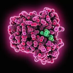
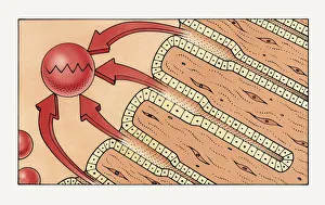
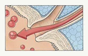
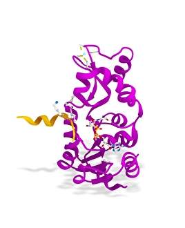
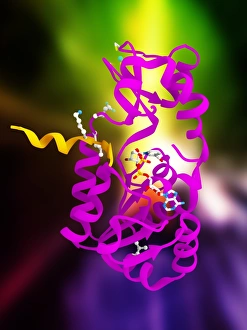
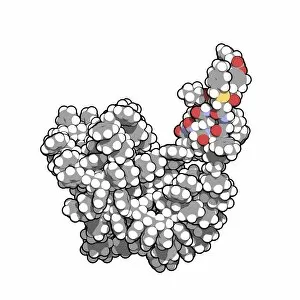
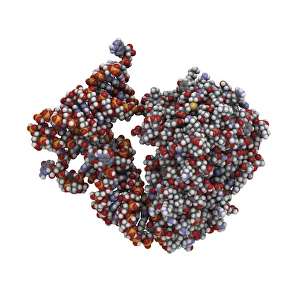
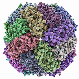
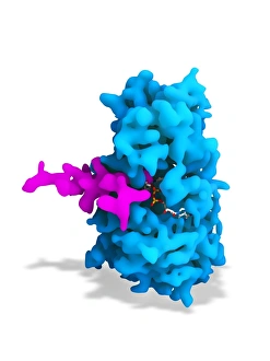
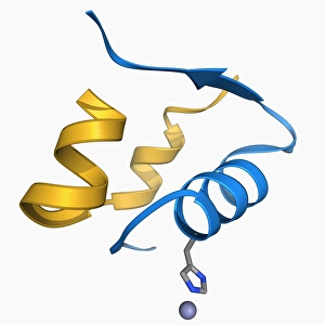
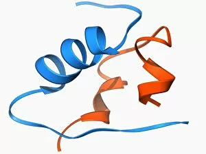
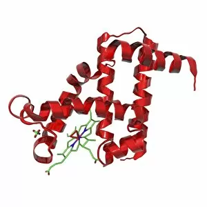
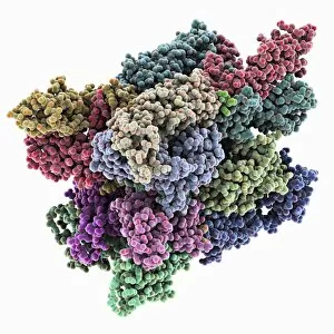
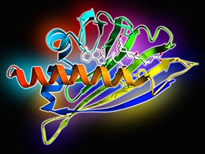
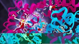
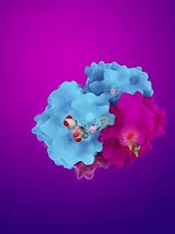
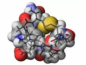
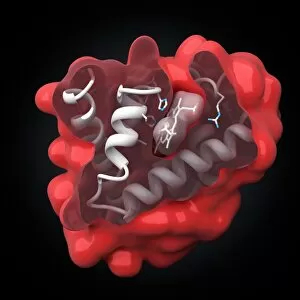
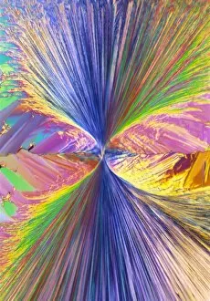
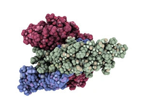
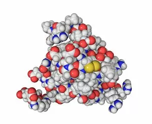
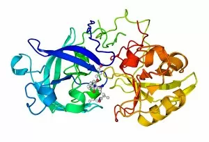
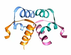
Peptides: Unveiling the Intricate World of Molecular Marvels DNA transcription and molecular model: Unlocking the secrets of life, DNA transcription orchestrates the synthesis of peptides, paving the way for countless biological wonders. Cone snail venom component molecule: Within the venomous embrace of a cone snail lies a tiny peptide molecule, holding immense potential for medical breakthroughs and pain management. Enkephalin crystals, light micrograph: Intricately arranged enkephalin crystals under microscopic scrutiny reveal nature's elegant design in these endogenous opioids that modulate our perception of pain. Gastric glands secreting pepsin illustration: Witness the fascinating process as gastric glands secrete pepsin to break down proteins into digestible peptides within our stomachs, fueling our bodies with vital nutrients. Pancreatic enzymes breaking down peptides illustration: Embark on a journey through human pancreatic ducts as enzymes diligently break down complex peptides into essential amino acids within the duodenum of our small intestine. Sirtuin enzyme and p53 artwork C017 / 3659: Marvel at this artistic representation showcasing the intricate dance between sirtuin enzyme and p53 protein, unraveling their role in cellular regulation and longevity. Insulin A chain molecule: Behold the elegance encapsulated within an insulin A chain molecule—a key player in regulating glucose metabolism—unleashing its power to maintain balance within our bodies. Transfer RNA-synthetase complex molecule: Delve into the realm where transfer RNA-synthetase complex molecules flawlessly match specific amino acids to their corresponding codons during protein synthesis—an exquisite feat of precision. Iron-containing protein molecular model: Discover how iron-containing proteins intricately bind this essential element, enabling crucial functions like oxygen transport or electron transfer throughout living organisms' systems.