Fraud Blocker Skip to main content

Proteins Collection (page 7)

Proteins: The Building Blocks of Life From the intricate network of nerve and glial cells to the mesmerizing patterns seen under a light micrograph

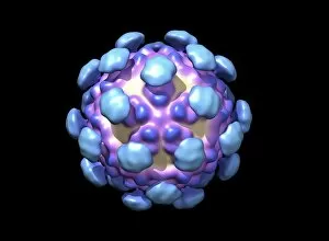
Background imageProteins Collection: Adenovirus particles, artwork C016 / 8964

Adenovirus particles, artwork C016 / 8964
Adenovirus particles. Computer artwork showing the external protein structure of adenovirus particles. Adenoviruses are the largest viruses not to have a protein coat covering their capsid
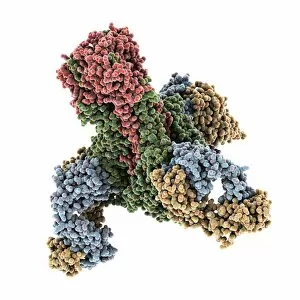
Background imageProteins Collection: Energy saving washing enzymes, concept C016 / 7514

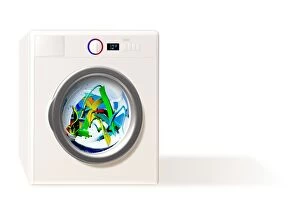
Energy saving washing enzymes, concept C016 / 7514
Energy saving washing enzymes, conceptual artwork

Background imageProteins Collection: Alpha-beta T-cell receptor

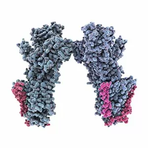
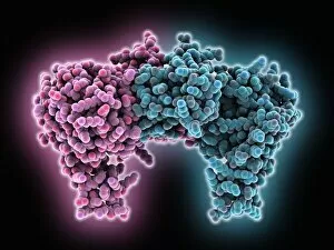
Alpha-beta T-cell receptor, molecular model. The T cell receptor (TCR) is a protein complex found on the surface of a type of white blood cell called T lymphocytes (or T cells)

Background imageProteins Collection: Transducin protein beta-gamma complex

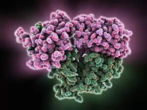
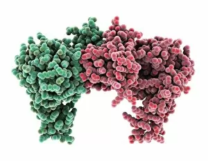
Transducin protein beta-gamma complex. Molecular model of the beta-gamma dimer of the heterotrimeric G protein transducin

Background imageProteins Collection: Nerve growth factor bound to receptor

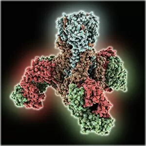
Nerve growth factor bound to receptor, molecular model. Nerve growth factor (NGF) complexed with the TrkA receptor. NGF is a neurotrophin that acts on the development and function of nerves

Background imageProteins Collection: Chromosome segregation protein

Chromosome segregation protein, molecular model. This proteins function is to aid the process of chromosome segregation during cell division and replication

Background imageProteins Collection: Nerve growth factor protein complex

Nerve growth factor protein complex, molecular model. This complex consists of nerve growth factor (NGF) in complex with four binding proteins

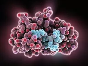
Background imageProteins Collection: Thymidylic acid-ribonuclease A complex

Thymidylic acid-ribonuclease A complex. Molecular model of a thymidylic acid tetramer (blue) in complex with ribonuclease A (red)

Background imageProteins Collection: Exosome complex, molecular model

Exosome complex, molecular model. This multi-protein complex functions to break up strands of RNA (ribonucleic acid, pink) during biochemical processes

Background imageProteins Collection: Bacterial ribosome and protein synthesis

Bacterial ribosome and protein synthesis. Molecular model showing a bacterial ribosome reading an mRNA (messenger ribonucleic acid) strand (blue) and synthesising a protein

Background imageProteins Collection: Tumour suppressor protein and DNA C016 / 6264

Tumour suppressor protein and DNA C016 / 6264
Tumour suppressor protein and DNA. Computer artwork showing a molecule of the tumour suppressor protein p53 (blue and green) bound to a strand of DNA (deoxyribonucleic acid, grey)

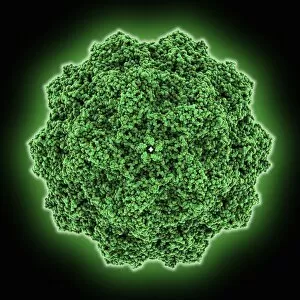
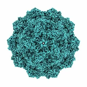
Background imageProteins Collection: Canine parvovirus capsid C015 / 8460

Canine parvovirus capsid C015 / 8460
Canine parvovirus capsid, molecular model. Canine parvovirus type 2 (CPV2) was first recognized in 1978 and spread worldwide in less than two years

Background imageProteins Collection: VDAC-1 ion channel protein C015 / 8250

VDAC-1 ion channel protein C015 / 8250
VDAC-1 ion channel protein, molecular model. This is the human voltage-dependent anion-selective channel protein 1 (VDAC-1)

Background imageProteins Collection: Streptavidin bacterial protein C015 / 8458

Streptavidin bacterial protein C015 / 8458
Streptavidin bacterial protein, molecular model. Streptavidin is a protein obtained from the bacterium Streptomyces avidinii

Background imageProteins Collection: Haemagglutinin viral surface protein C015 / 9965

Haemagglutinin viral surface protein C015 / 9965
Haemagglutinin viral surface protein. Molecular model of haemagglutinin, a surface protein from the influenza virus, complexed with a neutralising antibody

Background imageProteins Collection: Calcium ATPase ion pump C015 / 7143

Calcium ATPase ion pump C015 / 7143
Calcium ATPase ion pump, molecular model. This enzyme is found in muscle cell membranes, where it pumps calcium in and out of muscle cells and controls muscle contractions

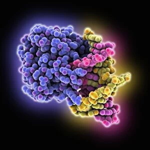
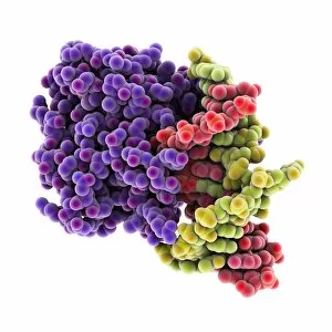
Background imageProteins Collection: DNA repair protein AlkB with DNA C016 / 0547

DNA repair protein AlkB with DNA C016 / 0547
DNA repair protein AlkB with DNA. Molecular model of the DNA (deoxyribonucleic acid) repair protein AlkB (blue) bound to a strand of double-stranded DNA (ds-DNA, pink and yellow)

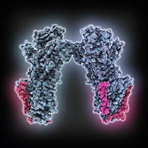
Background imageProteins Collection: Sodium-potassium ion pump proteins C015 / 9993

Sodium-potassium ion pump proteins C015 / 9993
Sodium-potassium ion pump proteins, molecular model. Sodium-potassium ATPase (adenosine triphosphatase) is an ATP-powered ion pump found in all animal cells

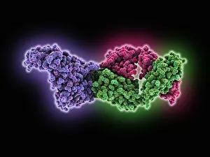
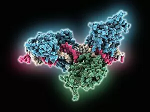
Background imageProteins Collection: Thrombin complexed with fibrinogen C015 / 7148

Thrombin complexed with fibrinogen C015 / 7148
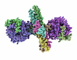
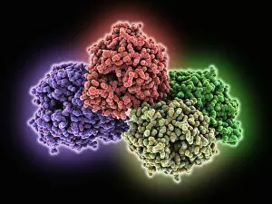
Thrombin complexed with fibrinogen, molecular model. The thrombin molecules (left and right, purple and green) are bound to the central part of the fibrinogen molecule (centre, multiple colours)

Background imageProteins Collection: 3-hydroxyacyl-CoA dehydrogenase C015 / 9940

3-hydroxyacyl-CoA dehydrogenase C015 / 9940
3-hydroxyacyl-CoA dehydrogenase, molecular model. This enzyme is found in human heart tissue, and catalyzes a reaction that is part of the beta-oxidation pathway

Background imageProteins Collection: Haemagglutinin viral surface protein C015 / 7124

Haemagglutinin viral surface protein C015 / 7124
Haemagglutinin viral surface protein. Molecular model of haemagglutinin, a surface protein from the influenza virus, complexed with a neutralising antibody

Background imageProteins Collection: Streptavidin bacterial protein C015 / 8457

Streptavidin bacterial protein C015 / 8457
Streptavidin bacterial protein, molecular model. Streptavidin is a protein obtained from the bacterium Streptomyces avidinii

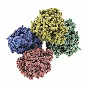
Background imageProteins Collection: Haemoglobin, molecular model C015 / 8938

Haemoglobin, molecular model C015 / 8938
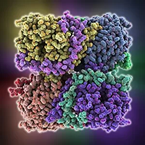
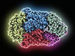
Haemoglobin, molecular model. This molecule transports oxygen around the body in red blood cells. It consists of four globin proteins (amino acid chains; orange, green, blue and purple)

Background imageProteins Collection: Sodium-potassium ion pump proteins C015 / 9997

Sodium-potassium ion pump proteins C015 / 9997
Sodium-potassium ion pump proteins, molecular model. Sodium-potassium ATPase (adenosine triphosphatase) is an ATP-powered ion pump found in all animal cells

Background imageProteins Collection: Retinal-producing oxygenase enzyme C015 / 7150

Retinal-producing oxygenase enzyme C015 / 7150
Retinal-producing oxygenase enzyme, molecular model. This enzyme, an oxygenase, plays a role in the production and metabolism of retinal and other apocarotenoids

Background imageProteins Collection: Haemagglutinin viral surface protein C015 / 9974

Haemagglutinin viral surface protein C015 / 9974
Haemagglutinin viral surface protein. Molecular model of haemagglutinin, a surface protein from the influenza virus, complexed with a neutralising antibody

Background imageProteins Collection: Central glycolytic gene regulator protein C016 / 0549

Central glycolytic gene regulator protein C016 / 0549
Central glycolytic gene regulator (CGGR) protein, molecular model. This protein binds to DNA (deoxyribonucleic acid) in the absence of glucose, blocking the transcription of certain genes

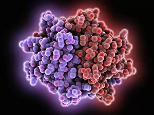
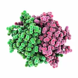
Background imageProteins Collection: Hemolysin-coregulated protein

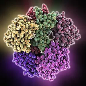
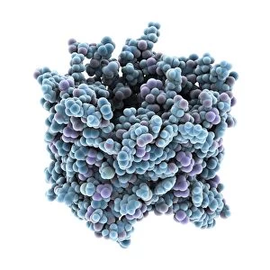
Hemolysin-coregulated protein, molecular model. This protein is from the bacterium Pseudomonas aeruginosa. It is a hexameric ring structure, named hcp1

Background imageProteins Collection: Canine parvovirus capsid C015 / 8459

Canine parvovirus capsid C015 / 8459
Canine parvovirus capsid, molecular model. Canine parvovirus type 2 (CPV2) was first recognized in 1978 and spread worldwide in less than two years

Background imageProteins Collection: DNA repair protein AlkB with DNA C016 / 0546

DNA repair protein AlkB with DNA C016 / 0546
DNA repair protein AlkB with DNA. Molecular model of the DNA (deoxyribonucleic acid) repair protein AlkB (purple) bound to a strand of double-stranded DNA (ds-DNA, red and green)

Background imageProteins Collection: Interferon-DNA transcription complex C015 / 8251

Interferon-DNA transcription complex C015 / 8251
Interferon-DNA transcription complex, molecular model. Bound to the DNA (deoxyribonucleic acid, green and yellow) is transcription factor p65, interferon regulatory factor 7

Background imageProteins Collection: Interferon-DNA transcription complex C015 / 8252

Interferon-DNA transcription complex C015 / 8252
Interferon-DNA transcription complex, molecular model. Bound to the DNA (deoxyribonucleic acid, pink and white) is transcription factor p65, interferon regulatory factor 7, interferon fusion protein

Background imageProteins Collection: Vitamin B12 import proteins C015 / 9942

Vitamin B12 import proteins C015 / 9942
Vitamin B12 import proteins, molecular model. This complex is the import proteins btuC, btuD, and btuF. The first two together form BtuCD

Background imageProteins Collection: Haemoglobin, molecular model C015 / 8939

Haemoglobin, molecular model C015 / 8939
Haemoglobin, molecular model. This molecule transports oxygen around the body in red blood cells. It consists of four globin proteins (amino acid chains; orange, green, blue and purple)

Background imageProteins Collection: VDAC-1 ion channel protein C015 / 8249

VDAC-1 ion channel protein C015 / 8249
VDAC-1 ion channel protein, molecular model. This is the human voltage-dependent anion-selective channel protein 1 (VDAC-1)

Background imageProteins Collection: Thrombin complexed with fibrinogen C015 / 7149

Thrombin complexed with fibrinogen C015 / 7149
Thrombin complexed with fibrinogen, molecular model. The thrombin molecules (left and right, brown and pink) are bound to the central part of the fibrinogen molecule (centre, multiple colours)

Background imageProteins Collection: Simian virus (SV40) large T antigen C015 / 7069

Simian virus (SV40) large T antigen C015 / 7069
Simian virus (SV40) large T antigen, molecular model. This antigen is from the simian vacuolating virus 40 (SV40). Large T antigens play a role in regulating the viral life cycle of

Background imageProteins Collection: Haemagglutinin viral surface protein C015 / 7123

Haemagglutinin viral surface protein C015 / 7123
Haemagglutinin viral surface protein. Molecular model of haemagglutinin, a surface protein from the influenza virus, complexed with a neutralising antibody

Background imageProteins Collection: 3-hydroxyacyl-CoA dehydrogenase C015 / 9941

3-hydroxyacyl-CoA dehydrogenase C015 / 9941
3-hydroxyacyl-CoA dehydrogenase, molecular model. This enzyme is found in human heart tissue, and catalyzes a reaction that is part of the beta-oxidation pathway

Background imageProteins Collection: Vitamin B12 import proteins C015 / 9943

Vitamin B12 import proteins C015 / 9943
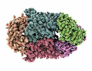
Vitamin B12 import proteins, molecular model. This complex is the import proteins btuC, btuD, and btuF. The first two together form BtuCD

Background imageProteins Collection: Calcium ATPase ion pump C015 / 7142

Calcium ATPase ion pump C015 / 7142
Calcium ATPase ion pump, molecular model. This enzyme is found in muscle cell membranes, where it pumps calcium in and out of muscle cells and controls muscle contractions

Background imageProteins Collection: Retinal-producing oxygenase enzyme C015 / 7151

Retinal-producing oxygenase enzyme C015 / 7151
Retinal-producing oxygenase enzyme, molecular model. This enzyme, an oxygenase, plays a role in the production and metabolism of retinal and other apocarotenoids

Background imageProteins Collection: Adeno-associated virus, molecular model C018 / 0449

Adeno-associated virus, molecular model C018 / 0449
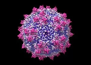
Adeno-associated virus (aV), molecular model. This image shows the core protein of the non-enveloped virus that surrounds the genetic material

Background imageProteins Collection: Hepatitis B virus, molecular model C018 / 0455

Hepatitis B virus, molecular model C018 / 0455
Hepatitis B virus core protein, molecular model. The virus causes hepatitis B, an inflammatory liver disease. The core proteins enclose the virus DNA and are in turn surrounded by a lipid envelope

Background imageProteins Collection: Norovirus capsid, molecular model C018 / 0457

Norovirus capsid, molecular model C018 / 0457
Norovirus capsid, molecular model. Also known as the winter vomiting bug, Noroviruses cause gastroenteritis and are highly contagious, infecting approximately 267 million people a year

Background imageProteins Collection: Astrovirus capsid, molecular model C018 / 0450

Astrovirus capsid, molecular model C018 / 0450
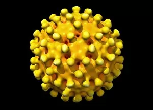
Astrovirus capsid, molecular model. This icosahedral virus was identified in 1975 using electron microscopy. It has a characteristic five-pointed symmetry to its surface, as seen here

Background imageProteins Collection: Hepatitis E virus, molecular model C018 / 0445

Hepatitis E virus, molecular model C018 / 0445
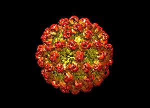
Hepatitis E virus core protein, molecular model. The virus causes hepatitis E, an inflammatory liver disease that usually only lasts a few weeks

Background imageProteins Collection: Human polio virus, molecular model

Human polio virus, molecular model
Human polio virus capsid, molecular model. Poliovirus causes poliomyelitis, a disease that can cause paralysis in up to 2 percent of patients, and in some cases death


All Professionally Made to Order for Quick Shipping

Proteins: The Building Blocks of Life From the intricate network of nerve and glial cells to the mesmerizing patterns seen under a light micrograph, proteins play an essential role in every aspect of our existence, and are like the conductors of our body's symphony, orchestrating vital processes that keep us alive and functioning. Take, for example, an anaesthetic inhibiting an ion channel C015 / 6718. Proteins act as gatekeepers, controlling what enters or exits our cells. In this case, they regulate the flow of ions necessary for transmitting nerve signals and maintaining proper cell function. But proteins don't just govern our internal workings; they also interact with external threats such as the avian flu virus. These microscopic invaders hijack host cells using their own protein machinery to replicate themselves. Understanding these interactions is crucial in developing effective treatments against viral infections. While some proteins protect us from harm, others contribute to overall well-being through a balanced diet. Our bodies require various types found in different foods to ensure optimal health and nutrition. The secondary structure is truly a work of art—a complex folding pattern that determines their shape and function. Artists have captured this beauty through stunning artwork showcasing these intricate molecular structures. One such structure is the nucleosome molecule—an elegant arrangement where DNA wraps around protein spools called histones—forming compact units within chromosomes. This organization allows efficient storage and retrieval of genetic information during cell division or gene expression. Antibodies are another remarkable class depicted in captivating artwork. These specialized molecules recognize foreign substances like bacteria or viruses and neutralize them by binding tightly to specific targets on their surface—an extraordinary defense mechanism employed by our immune system. Speaking of bacteria, their ribosomes serve as factories producing new proteins based on instructions encoded in DNA—the blueprint for life itself. Understanding bacterial ribosomes has led to groundbreaking discoveries in antibiotic development, combating infectious diseases that threaten human health.