
Tumor Collection
DDE-90026224, DDE-90026223, DDE-90026222, DDE-90026221, DDE-90026220, DDE-90026219


All Professionally Made to Order for Quick Shipping
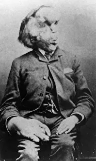
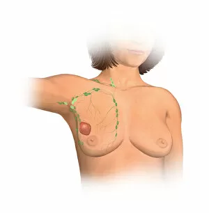
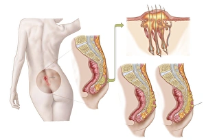
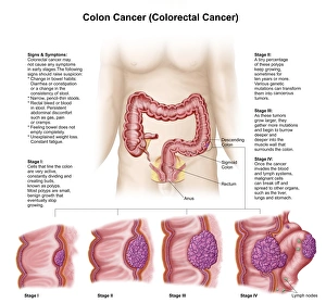
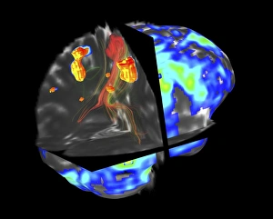
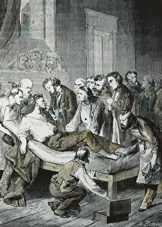
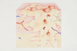
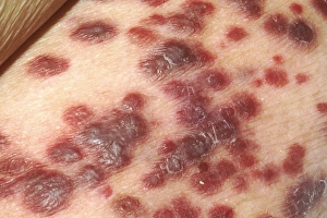
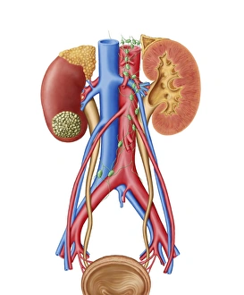
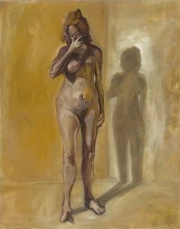
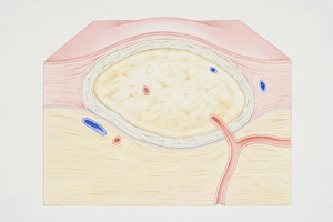
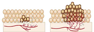
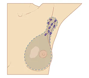
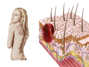
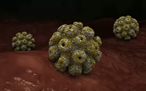
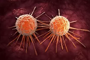
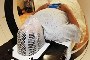
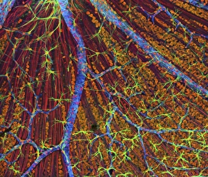
DDE-90026224, DDE-90026223, DDE-90026222, DDE-90026221, DDE-90026220, DDE-90026219, and DDE-90026218 - these are the codes that represent a battle fought by countless individuals against the silent enemy within: tumors. In 1889, Joseph Carey Merrick's haunting black and white photo captured not just his physical appearance but also the resilience of those facing unimaginable challenges. His story reminds us that behind every tumor lies a person with dreams and aspirations. An anterior view of female anatomy reveals breast tissue marred by a tumor. This image serves as a stark reminder of the importance of early detection and regular check-ups to safeguard our health. William Thomas Green Morton was an American pioneer in anesthesia who tragically lost his life at just 49 years old. His legacy lives on as medical professionals continue to explore innovative ways to combat tumors and improve patient outcomes. A medical illustration depicting a pilonidal cyst near the natal cleft serves as a visual representation of how even seemingly innocuous areas can be affected by tumors. It highlights the need for vigilance in monitoring our bodies for any signs or symptoms. Colon cancer is one of the most prevalent forms of this disease worldwide. A striking medical illustration showcases its different stages - from initial growth to advanced malignancy - urging us all to prioritize screenings and adopt healthy lifestyle choices. Tumors may be formidable foes, but they cannot extinguish hope or determination. Let us stand together in solidarity with those battling these insidious growths while supporting research efforts aimed at finding effective treatments and ultimately eradicating them altogether.