
Ischium Collection
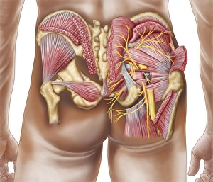
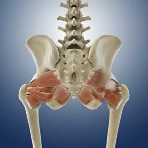
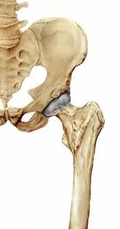
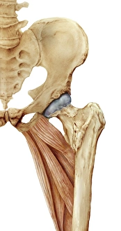
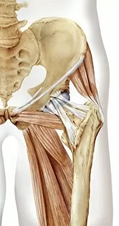
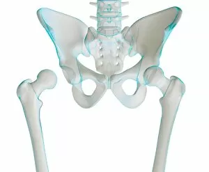
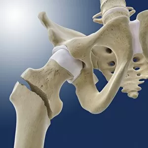
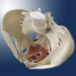
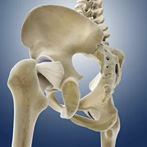
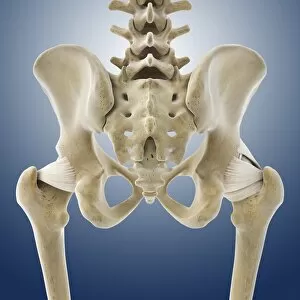
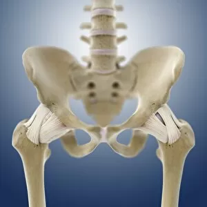
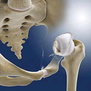
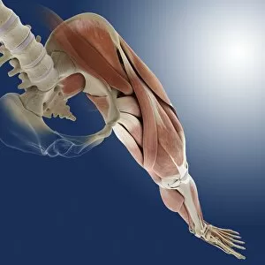
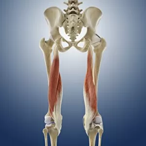
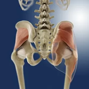
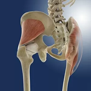
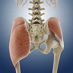
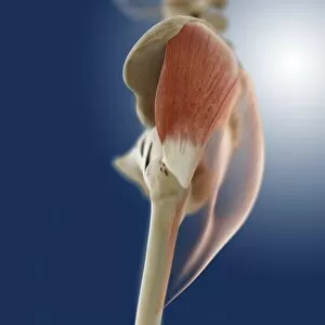
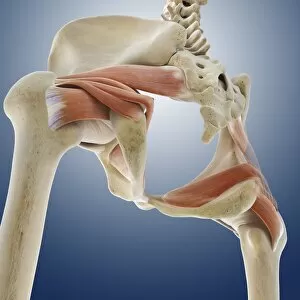
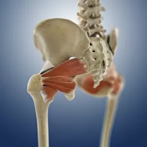
The ischium, a vital component of the gluteal muscles in the human buttocks, plays a crucial role in our body's support and movement


All Professionally Made to Order for Quick Shipping
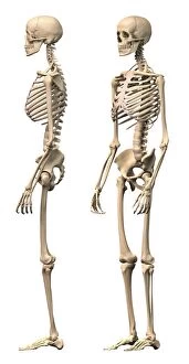
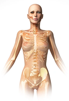
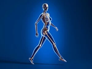
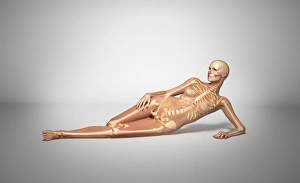
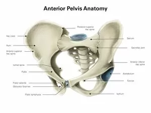
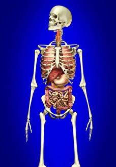
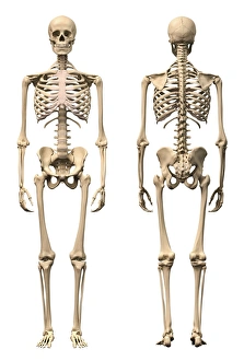
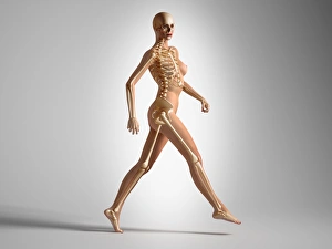
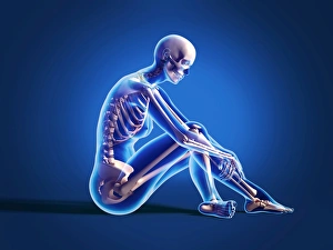
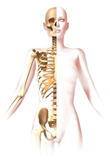
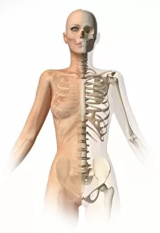
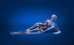
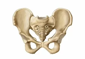
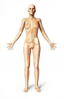
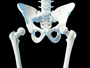
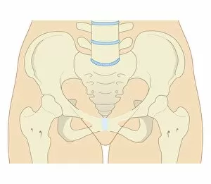
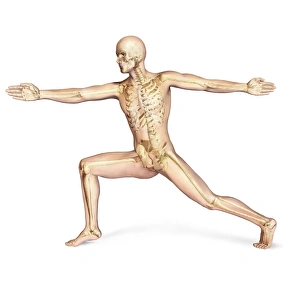
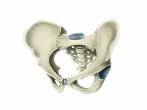
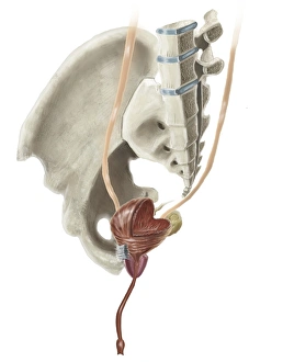
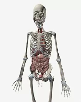
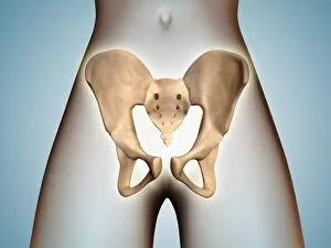
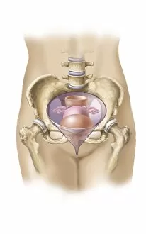
The ischium, a vital component of the gluteal muscles in the human buttocks, plays a crucial role in our body's support and movement. Whether viewed from the side or perspective angle, its presence is undeniable in both male and female anatomies. In an intriguing juxtaposition, a naked woman lies down with skeletal bones superimposed upon her body, revealing the intricate connection between internal organs and this essential bone structure. From an anterior view of the pelvis to a male skeleton against a blue background showcasing internal organs, we witness how the ischium harmoniously integrates within our bodies' framework. X-ray views further emphasize its significance - whether it be a woman sitting on the floor or walking confidently - as skeletal bones are overlaid onto their forms. Even through 3D renderings, where realism meets artistry, we observe how this bone contributes to balance and stability during motion. With artwork depicting hip joint bones and anatomy serving as visual aids (artwork C014 / 2032), we gain deeper insight into this remarkable element of our physiology that often goes unnoticed beneath our skin's surface.